Concept explainers
The accompanying pedigree below shows a family in which an autosomal recessive disorder is presents. Family members
Excluding
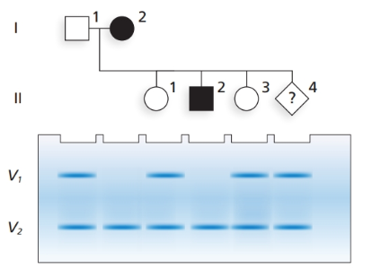
What is the genotype of each family member, including
What are the synthetic disease gene and VNTR alleles in
What is the chance
Want to see the full answer?
Check out a sample textbook solutionChapter 5 Solutions
Genetic Analysis: An Integrated Approach (3rd Edition)
- A couple was referred for genetic counseling because they wanted to know the chances of having a child with dwarfism. Both the man and the woman had achondroplasia (MIM 100800), the most common form of short-limbed dwarfism. The couple knew that this condition is inherited as an autosomal dominant trait, but they were unsure what kind of physical manifestations a child would have if it inherited both mutant alleles. They were each heterozygous for the FGFR3 (MIM 134934) allele that causes achondroplasia. Normally, the protein encoded by this gene interacts with growth factors outside the cell and receives signals that control growth and development. In achrodroplasia, a mutation alters the activity of the receptor, resulting in a characteristic form of dwarfism. Because both the normal and mutant forms of the FGFR3 protein act before birth, no treatment for achrondroplasia is available. The parents each carry one normal allele and one mutant allele of FGRF3, and they wanted information on their chances of having a homozygous child. The counsellor briefly reviewed the phenotypic features of individuals with achondroplasia. These include facial features (large head with prominent forehead; small, flat nasal bridge; and prominent jaw), very short stature, and shortening of the arms and legs. Physical examination and skeletal X-ray films are used to diagnose this condition. Final adult height is approximately 4 feet. Because achondroplasia is an autosomal dominant condition, a heterozygote has a 1-in-2, or 50%, chance of passing this trait to his or her offspring. However, about 75% of those with achondroplasia have parents of average size who do not carry the mutant allele. In these cases, achondroplasia is due to a new mutation. In the couple being counseled, each individual is heterozygous, and they are at risk for having a homozygous child with two copies of the mutated gene. Infants with homozygous achondroplasia are either stillborn or die shortly after birth. The counselor recommended prenatal diagnosis via ultrasounds at various stages of development. In addition, a DNA test is available to detect the homozygous condition prenatally. Should the parents be concerned about the heterozygous condition as well as the homozygous mutant condition?arrow_forwardA couple was referred for genetic counseling because they wanted to know the chances of having a child with dwarfism. Both the man and the woman had achondroplasia (MIM 100800), the most common form of short-limbed dwarfism. The couple knew that this condition is inherited as an autosomal dominant trait, but they were unsure what kind of physical manifestations a child would have if it inherited both mutant alleles. They were each heterozygous for the FGFR3 (MIM 134934) allele that causes achondroplasia. Normally, the protein encoded by this gene interacts with growth factors outside the cell and receives signals that control growth and development. In achrodroplasia, a mutation alters the activity of the receptor, resulting in a characteristic form of dwarfism. Because both the normal and mutant forms of the FGFR3 protein act before birth, no treatment for achrondroplasia is available. The parents each carry one normal allele and one mutant allele of FGRF3, and they wanted information on their chances of having a homozygous child. The counsellor briefly reviewed the phenotypic features of individuals with achondroplasia. These include facial features (large head with prominent forehead; small, flat nasal bridge; and prominent jaw), very short stature, and shortening of the arms and legs. Physical examination and skeletal X-ray films are used to diagnose this condition. Final adult height is approximately 4 feet. Because achondroplasia is an autosomal dominant condition, a heterozygote has a 1-in-2, or 50%, chance of passing this trait to his or her offspring. However, about 75% of those with achondroplasia have parents of average size who do not carry the mutant allele. In these cases, achondroplasia is due to a new mutation. In the couple being counseled, each individual is heterozygous, and they are at risk for having a homozygous child with two copies of the mutated gene. Infants with homozygous achondroplasia are either stillborn or die shortly after birth. The counselor recommended prenatal diagnosis via ultrasounds at various stages of development. In addition, a DNA test is available to detect the homozygous condition prenatally. What is the chance that this couple will have a child with two copies of the dominant mutant gene? What is the chance that the child will have normal height?arrow_forwardA proband female with an unidentified disease seeks the advice of a genetic counselor before starting a family. Based on the following data, the counselor constructs a pedigree encompassing three generations: (1) The maternal grandfather of the proband has the disease. (2) The mother of the proband is unaffected and is the youngest of five children, the three oldest being male. (3) The proband has an affected older sister, but the youngest siblings are unaffected twins (boy and girl). (4) All the individuals who have the disease have been revealed. Duplicate the counselors featarrow_forward
- The pedigree shows inheritance of the autosomal recessive trait cystic fibrosis in a family. Family members who are known to have the disease are represented by black shading. I 1 2 3 4 II 1 3 4 6 8 7 III 1 2 3 4 5 6 7 Part A: Select the best answer from the boxes below to indicate the likely genotypes of the following individuals, where F indicates the wild-type (usual) allele and f indicates the cystic fibrosis allele. The first part of their number (I, II or III) indicates their generation or row, whilst the second part of their number (1-8) indicates the individual within that generation or row. |-4 Il-3 Il-8 III-3arrow_forwardThere are six types of agglutinogen named C,D, E and c,d,e.the first three are dominant and last three are recessive.discussarrow_forwardPedigree attached shows an autosomal recessive genetic disease. G is the normal allele and g is the disease-causing allele. Individual 1’s father is heterozygous (*) and his mother is homozygous dominant. Other individuals in the pedigree may be carriers, but are not marked. The question mark (?) indicates that you do not yet know anything about this individual’s phenotype with regard to the disease. part a) What is the probability that individuals 1 and 2 will have a child (5) who is a boy with the disease (the child is unborn and the sex is not yet known)? a)1/8 b)1/4 c)0 d)1/16 part b) What is the probability that the daughter (6) that individual 3 and 4 just had will have the disease? a)1/8 b)1/6 c)1/4 d)1/12arrow_forward
- The pedigree below shows the phenotypes of the ABO blood groups and Rhesus factors [positive (+) and negative (-)] for several members of a family. I (B+ AB- 1 2 3 4 II O- A+ В- B- AB+ A+ 1 2 4 5 6 a. What are the ABO blood group genotypes of individuals I-1 and I-2? b. Which child/ren of individual I-4 can donate blood to him? c. Which individual in the pedigree can donate blood to all the other individuals in the pedigree?arrow_forwardThe pedigree shows inheritance of an autosomal recessive disease in an extended family. Assume unrelated individuals marrying into the family do not carry the disease, unless there is reason to believe otherwise. What is the chance that IV-3 and IV-4 will have a child with the disease? Individuals I-1, Il-5, III-5 and III-16 have the disease. 2 1 7. III-18 2 3 5 6 17 8. 9 10 11 12 13 14 15 16 17 III-19 IV IV-3 IV-4 IV-5 IV-6 IV-1 IV-2 O a. 1/8 b. 1/12 C. 1/16 d. 3/16 e. 1/24 f. 1/32 g. 3/32 h. 1/64 FEB 17 MacBook Air 6, ... 5. %Darrow_forwardBelow is a pedigree chart for a family that has a history of Alkaptonuria. Individuals infected with this condition can have darkened skin, brown urine, and can suffer from joint damage and other complications. Given this pedigree answer the following questions. Given the data in the pedigree chart is this genetic condition autosomal dominant or autosomal recessive? What are the genotypes for #1, #2, and #3? If either of the 4th generation "aa" females were to mate with a homozygous dominant male would any of their offspring illustrate the phenotype? Why or why not?arrow_forward
- In this case a family history revealed a genetic basis for the disorder. The pedigree is shown in Fig. 1 Below. Key Ø Female: affected Female: unaffected || IV V 5600 orize 077808 15 10 9 10 CHO વ Male: affected Male: unaffected Deceased Disease status not given Dizygotic twins Monozygotic twins Fig. 1 Disease pedigree. Five generations I, II, III, IV, V are shown. Females are represented by circles, males by squares, dizygotic (non-identical) twins by diagonal lines originating from the same point, Monozygotic (identical) twins by diagonal lines originating from the same point and joined symbols and deceased by a diagonal line through the symbol. Filled symbols indicate that the individual displays the disease phenotype. Unfilled symbols indicate that the individual does not display the disease phenotype. Carriers of the disease are not indicated. Information on disease status is not known for generation I and is omitted for the individuals represented by a symbol with an asterisk.…arrow_forwardPlease consider the following pedigree. Assume that people who marry in to the family do not carry the allele. Assume complete penetrance. I II III 3 IV 1 2 a. Is it possible for the inheritance pattern for the trait illustrated in this pedigree to be as a result of each of the following? Answer yes or no. (i) an autosomal recessive allele (AR) (ii) an autosomal dominant allele (AD) (iii) a X-linked recessive allele (XR) (iv) a X-linked dominant allele (XD) b. Based strictly on the characteristic patterns of inheritance that define the four different options in (a), give a definitive motivation for the most likely mode of inhertance.arrow_forwardBoth hemophilia (h) and favism (gd) are inherited as X-linked recessive traits. Hemophilia is an inherited disorder of blood clotting, and favism is an inherited hemolytic anemia caused by absence of the enzyme glucose-6-phosphate dehydrogenase. A phenotypically normal woman is known to have the X chromosome genotype h + / + gd. The frequency of recombination between h and gd is 16%. What proportion of sons born to this woman are expected to be phenotypically normal with respect to both hemophilia and favism?arrow_forward
- Human Heredity: Principles and Issues (MindTap Co...BiologyISBN:9781305251052Author:Michael CummingsPublisher:Cengage Learning