3. A student in my lab was trying to express the cytosolic (not membrane bound) enzyme PseG and decided to express the enzyme as a His fusion (PseG-His) to ease purification. In his initial experiments, he (1) expressed the protein in bacterial cells (E. coli), (2) lysed the bacteria using a gentle lysis buffer (one that would solubilize folded proteins), and (3) assessed the protein's presence in whole-cells (WC) pre-lysis, and pellet (P) versus soluble (S) cell fractions post-lysis, by SDS-PAGE analysis. In his first expression experiment, he collected the data shown in Figure 1: kD 58 46 kD MW WC PP SS 175 80 30- 25 10- Coomassie-stained protein gel a. From these initial data, what can you conclude about the following features of PseG-His6 in E. coli? Expressed? Soluble? Folded? 10 In a follow-up experiment, my student (1) expressed the protein in bacterial cells (E. coli), (2) lysed the bacteria using a harsh lysis buffer that included 6 M urea (a detergent), and (3) assessed the protein's presence in whole-cells (WC) pre-lysis, and pellet (P) versus soluble (S) cell fractions post-lysis, by SDS- PAGE analysis. In this second experiment, he collected the data shown in Figure 2: ← PseG-His6 fusion WC PS expression was Figure 1. Protein experiment #1. PseG-His6 expressed in E. coli. E. coli were then burst open using a gentle lysis buffer that would solubilize folded proteins. The presence of PseG-Hise in whole- cells (WC) before lysis, and pellet (P) and supernatant (S) fractions post-lysis, was assessed by SDS-PAGE analysis. Pellet and supernatant fractions were run in duplicate. ← PseG-His6 fusion Figure 2. Protein expression experiment #2. PseG-His6 was expressed in E. coli. E. coli were then burst open using a harsh lysis buffer that included 6 M urea (a detergent). The presence of PseG-Hise in whole-cells (WC) before lysis, and pellet (P) and supernatant (S) fractions post-lysis, was assessed by SDS-PAGE analysis.
3. A student in my lab was trying to express the cytosolic (not membrane bound) enzyme PseG and decided to express the enzyme as a His fusion (PseG-His) to ease purification. In his initial experiments, he (1) expressed the protein in bacterial cells (E. coli), (2) lysed the bacteria using a gentle lysis buffer (one that would solubilize folded proteins), and (3) assessed the protein's presence in whole-cells (WC) pre-lysis, and pellet (P) versus soluble (S) cell fractions post-lysis, by SDS-PAGE analysis. In his first expression experiment, he collected the data shown in Figure 1: kD 58 46 kD MW WC PP SS 175 80 30- 25 10- Coomassie-stained protein gel a. From these initial data, what can you conclude about the following features of PseG-His6 in E. coli? Expressed? Soluble? Folded? 10 In a follow-up experiment, my student (1) expressed the protein in bacterial cells (E. coli), (2) lysed the bacteria using a harsh lysis buffer that included 6 M urea (a detergent), and (3) assessed the protein's presence in whole-cells (WC) pre-lysis, and pellet (P) versus soluble (S) cell fractions post-lysis, by SDS- PAGE analysis. In this second experiment, he collected the data shown in Figure 2: ← PseG-His6 fusion WC PS expression was Figure 1. Protein experiment #1. PseG-His6 expressed in E. coli. E. coli were then burst open using a gentle lysis buffer that would solubilize folded proteins. The presence of PseG-Hise in whole- cells (WC) before lysis, and pellet (P) and supernatant (S) fractions post-lysis, was assessed by SDS-PAGE analysis. Pellet and supernatant fractions were run in duplicate. ← PseG-His6 fusion Figure 2. Protein expression experiment #2. PseG-His6 was expressed in E. coli. E. coli were then burst open using a harsh lysis buffer that included 6 M urea (a detergent). The presence of PseG-Hise in whole-cells (WC) before lysis, and pellet (P) and supernatant (S) fractions post-lysis, was assessed by SDS-PAGE analysis.
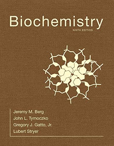
Biochemistry
9th Edition
ISBN:9781319114671
Author:Lubert Stryer, Jeremy M. Berg, John L. Tymoczko, Gregory J. Gatto Jr.
Publisher:Lubert Stryer, Jeremy M. Berg, John L. Tymoczko, Gregory J. Gatto Jr.
Chapter1: Biochemistry: An Evolving Science
Section: Chapter Questions
Problem 1P
Related questions
Question

Transcribed Image Text:3. A student in my lab was trying to express the cytosolic (not membrane bound) enzyme PseG and decided to
express the enzyme as a Hisí fusion (PseG-His) to ease purification. In his initial experiments, he (1) expressed
the protein in bacterial cells (E. coli), (2) lysed the bacteria using a gentle lysis buffer (one that would solubilize
folded proteins), and (3) assessed the protein's presence in whole-cells (WC) pre-lysis, and pellet (P) versus
soluble (S) cell fractions post-lysis, by SDS-PAGE analysis. In his first expression experiment, he collected the
data shown in Figure 1:
kD
58
46
kD MW WC PP
175
80
58
46
30
25
│I
T||| || |
10-
Coomassie-stained protein gel
a. From these initial data, what can you conclude about the following features of PseG-His6 in E. coli?
Expressed?
Soluble?
Folded?
10->>
In a follow-up experiment, my student (1) expressed the protein in bacterial cells (E. coli), (2) lysed the
bacteria using a harsh lysis buffer that included 6 M urea (a detergent), and (3) assessed the protein's
presence in whole-cells (WC) pre-lysis, and pellet (P) versus soluble (S) cell fractions post-lysis, by SDS-
PAGE analysis. In this second experiment, he collected the data shown in Figure 2:
SS
WC PS
← PseG-His6
fusion
Coomassie-stained protein gel
expression
was
Figure 1. Protein
experiment #1. PseG-His6
expressed in E. coli. E. coli were then
burst open using a gentle lysis buffer
that would solubilize folded proteins.
The presence of PseG-His6 in whole-
cells (WC) before lysis, and pellet (P)
and supernatant (S) fractions_post-lysis,
was assessed by SDS-PAGE analysis.
Pellet and supernatant fractions were
run in duplicate.
PseG-His6
fusion
Figure 2. Protein expression experiment
#2. PseG-His6 was expressed in E. coli. E.
coli were then burst open using a harsh
lysis buffer_that included 6 M urea (a
detergent). The presence of PseG-His in
whole-cells (WC) before lysis, and pellet (P)
and supernatant (S) fractions post-lysis, was
assessed by SDS-PAGE analysis.
![b. What can you conclude about the following features of PseG-His。 in E. coli in this follow-up
experiment?
Next, my student set out to purify the PseG-His present in the supernatant from the harsh lysis (obtained
in Figure 2). He loaded the lysate onto a Ni+² column, rinsed the column with buffer containing increasing
concentrations of imidazole, and analyzed fractions for the presence of PseG-His6:
C.
kD
30
25
||
Expressed?
Soluble?
Folded?
10->
[imidizole]
50-100 mM
200 mM imidizole
PseG-His
fusion
Figure 3. Fractions from a Ni+²-column
analyzed by SDS-PAGE. The column was
loaded with E. coli lysate and subsequently
rinsed with buffer containing increasing
concentrations of imidazole as indicated.
imidazole
Coomassie-stained protein gel
What type of purification is this? What happened when 200 mM imidazole was added to the column?
Why?
d. Is the purified protein obtained in Figure 3 active? If not, what could my student do to help this protein
gain activity?](/v2/_next/image?url=https%3A%2F%2Fcontent.bartleby.com%2Fqna-images%2Fquestion%2Fdc99cc4f-9438-44d8-9b43-49724a6b39cf%2F18c00587-16a0-46f9-9a77-67b4ac28a011%2Fhaqsm5_processed.png&w=3840&q=75)
Transcribed Image Text:b. What can you conclude about the following features of PseG-His。 in E. coli in this follow-up
experiment?
Next, my student set out to purify the PseG-His present in the supernatant from the harsh lysis (obtained
in Figure 2). He loaded the lysate onto a Ni+² column, rinsed the column with buffer containing increasing
concentrations of imidazole, and analyzed fractions for the presence of PseG-His6:
C.
kD
30
25
||
Expressed?
Soluble?
Folded?
10->
[imidizole]
50-100 mM
200 mM imidizole
PseG-His
fusion
Figure 3. Fractions from a Ni+²-column
analyzed by SDS-PAGE. The column was
loaded with E. coli lysate and subsequently
rinsed with buffer containing increasing
concentrations of imidazole as indicated.
imidazole
Coomassie-stained protein gel
What type of purification is this? What happened when 200 mM imidazole was added to the column?
Why?
d. Is the purified protein obtained in Figure 3 active? If not, what could my student do to help this protein
gain activity?
Expert Solution
Step 1
The expression of proteins in a prokaryotic or eukaryotic cell involves transcription and translation progress of the gene of interest n the cell in which the protein is being expressed.
The bacterial expression system can act as rapid and simple systems of expressing large amounts of recombinant eukaryotic proteins.
However some recombinant proteins however tend to form inclusion bodies upon their expression in bacterial cells.
Step by step
Solved in 2 steps

Recommended textbooks for you

Biochemistry
Biochemistry
ISBN:
9781319114671
Author:
Lubert Stryer, Jeremy M. Berg, John L. Tymoczko, Gregory J. Gatto Jr.
Publisher:
W. H. Freeman

Lehninger Principles of Biochemistry
Biochemistry
ISBN:
9781464126116
Author:
David L. Nelson, Michael M. Cox
Publisher:
W. H. Freeman

Fundamentals of Biochemistry: Life at the Molecul…
Biochemistry
ISBN:
9781118918401
Author:
Donald Voet, Judith G. Voet, Charlotte W. Pratt
Publisher:
WILEY

Biochemistry
Biochemistry
ISBN:
9781319114671
Author:
Lubert Stryer, Jeremy M. Berg, John L. Tymoczko, Gregory J. Gatto Jr.
Publisher:
W. H. Freeman

Lehninger Principles of Biochemistry
Biochemistry
ISBN:
9781464126116
Author:
David L. Nelson, Michael M. Cox
Publisher:
W. H. Freeman

Fundamentals of Biochemistry: Life at the Molecul…
Biochemistry
ISBN:
9781118918401
Author:
Donald Voet, Judith G. Voet, Charlotte W. Pratt
Publisher:
WILEY

Biochemistry
Biochemistry
ISBN:
9781305961135
Author:
Mary K. Campbell, Shawn O. Farrell, Owen M. McDougal
Publisher:
Cengage Learning

Biochemistry
Biochemistry
ISBN:
9781305577206
Author:
Reginald H. Garrett, Charles M. Grisham
Publisher:
Cengage Learning

Fundamentals of General, Organic, and Biological …
Biochemistry
ISBN:
9780134015187
Author:
John E. McMurry, David S. Ballantine, Carl A. Hoeger, Virginia E. Peterson
Publisher:
PEARSON