Concept explainers
Videos
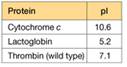
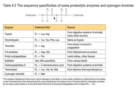
You are a summer intern in a clinical hematology lab. The lab director gives you a sample of a patient's blood proteins and asks you to characterize the thrombin in the sample. She also tells you that thrombin is a serine protease important in blood clotting (see Table 5.3), and this patient is a newborn with uncontrolled bleeding.
a. To characterize the thrombin in the sample, you must remove two proteins that interfere with the thrombin activity assay: cytochrome c and lactoglobin. You find some CM-cellulose (see Figure 5A.5) and a phosphate buffer (pH 6.4) on the shelf in your lab. You decide to load the protein sample onto a column of CM-cellulose equilibrated in the pH = 6.4 buffer. Predict the order of elution for the three proteins shown in the accompanying table. At pH = 6.4, which protein(s) do you predict will remain bound to the column?
b. List two different ways you could change the buffer to elute the bound protein(s) and achieve proper separation of the proteins.
c. You are surprised to observe that the patient's thrombin flows through the CM-cellulose column at pH = 6.4 and does not bind. Confident in your technique, you suspect the patient's thrombin is different from wild-type thrombin. Using a different buffer system, you manage to purify some of the patient's thrombin, and you submit the purified sample for amino acid sequencing. The sequence analysis shows that the patient's thrombin contains a mutation in the enzyme active site. A lysine residue in the wild type has been mutated to an asparagine in the patient's thrombin. Does this mutation explain the anomalous CM- cellulose binding behavior you observed?
d. How many
e. Based on their side-chain structures, compare and contrast the potential of Lys and Asn to form noncovalent interactions. In other words, can each form H bonds and/or salt bridges and/or van der Waals contacts?
Want to see the full answer?
Check out a sample textbook solution
Chapter 5 Solutions
Biochemistry: Concepts and Connections (2nd Edition)


- Draw the predominant form of glutamic acid at pH = 8.4. The pKa of the side chain is 4.1. Include proper stereochemistry. HO H2N OH pH = 8.4arrow_forwardHow would I draw this?arrow_forwardCalculate the standard change in Gibbs free energy, AGrxn, for the given reaction at 25.0 °C. Consult the table of thermodynamic properties for standard Gibbs free energy of formation values. NH,Cl(s) →NH; (aq) + C1 (aq) AGrxn -7.67 Correct Answer Determine the concentration of NH+ (aq) if the change in Gibbs free energy, AGrxn, for the reaction is -9.27 kJ/mol. 6.49 [NH+] Incorrect Answer kJ/mol Marrow_forward
- What are some topics of interest that neurotoxicologists study? For example, toxin-induced seizures, brain death, and such along those lines?arrow_forwardCould you help me with the explanation of the answer to exercise 15, chapter 1 of Lehinger Question Nombramiento de estereoisómeros con dos carbonos quirales utilizando el sistema RS(R,R)El isómero del metilfenidato (Ritalin) se utiliza para tratar el trastorno por déficit de atención con hiperactividad (TDAH).(S,S)El isómero es un antidepresivo. Identifique los dos carbonos quirales en la siguiente estructura. ¿Es este el(R,R)o el(S,S)¿isómero? Dibuja el otro isómero. Nombramiento de estereoisómeros con dos carbonos quirales utilizando el sistema RS(R,R)El isómero del metilfenidato (Ritalin) se utiliza para tratar el trastorno por déficit de atención con hiperactividad (TDAH).(S,S)El isómero es un antidepresivo.arrow_forwardThe reaction A+B → C + D AG°' = -7.3 kcal/mol can be coupled with which of the following unfavorable reactions to drive it forward? A. EFG+HAG° = 5.6 kcal/mol. B. J+KZ+A AG° = 2.3 kcal/mol. C. P+RY+DAG° = 8.2 kcal/mol. D. C + T → V + W AG°' = -5.9 kcal/mol. E. AN→ Q+KAG°' = 4.3 kcal/mol.arrow_forward
- What would be the toxicological endpoints for neurotoxicity?arrow_forwardWhat are "endpoints" in toxicology exactly? Please give an intuitive easy explanationarrow_forwardFura-2 Fluorescence (Arbitrary Unit) 4500 4000 3500 3000 2500 2000 1500 1000 500 [Ca2+]=2970nM, 25°C [Ca2+] 2970nM, 4°C [Ca2+]=0.9nM, 25°C [Ca2+] = 0.9nM, 4°C 0 260 280 300 340 360 380 400 420 440 Wavelength (nm) ← < The figure on the LHS shows the excitation spectra of Fura-2 (Em = 510 nm) in 2 solutions with two different Ca2+ ion concentration as indicated. Except for temperature, the setting for excitation & signal acquisition was identical.< ப a) The unit in Y-axis is arbitrary (unspecified). Why? < < b) Compare & contrast the excitation wavelength of the Isosbestic Point of Fura-2 at 25 °C & 4 °C. Give a possible reason for the discrepancy. < c) The fluorescence intensity at 25 °C & 4 °C are different. Explain why with the concept of electronic configuration. <arrow_forward

 Human Heredity: Principles and Issues (MindTap Co...BiologyISBN:9781305251052Author:Michael CummingsPublisher:Cengage Learning
 Principles Of Radiographic Imaging: An Art And A ...Health & NutritionISBN:9781337711067Author:Richard R. Carlton, Arlene M. Adler, Vesna BalacPublisher:Cengage Learning
 Human Physiology: From Cells to Systems (MindTap ...BiologyISBN:9781285866932Author:Lauralee SherwoodPublisher:Cengage Learning
 Anatomy & PhysiologyBiologyISBN:9781938168130Author:Kelly A. Young, James A. Wise, Peter DeSaix, Dean H. Kruse, Brandon Poe, Eddie Johnson, Jody E. Johnson, Oksana Korol, J. Gordon Betts, Mark WomblePublisher:OpenStax College