Refer to the attached DNA model. In the spaces provided on the attached table, identify the nitrogenous bases for the first 5 base pairs from the top, and for the 5 base pairs from the bottom of the model. Use ‘A’, ‘G’, ‘C’, ‘T’ to indicate the proper base. Polarity does matter.


DNA and RNA
Deoxyribonucleic acid (DNA) is usually called the blueprint of life. Deoxyribose is a monosaccharide that has a key function in the synthesis of deoxyribonucleic acid. One less oxygen-containing hydroxyl group occurs in deoxyribose sugar. Nucleic acid, deoxyribonucleic acid, is one of the natural components. Deoxyribonucleic acid is a double-stranded molecule. Watson and Crick postulated the double-stranded model of the helix. A deoxyribonucleic acid is a molecular group that carries and transmits genetic information from parents to offspring. All eukaryotic and prokaryotic cells are involved.
DNA as the Genetic Material
DNA, or deoxyribonucleic acid, is a long polymeric nucleic acid molecule discovered in the late 1930s. It is a polymer; a long chain-like molecule made up of several monomers connected in a sequence. It possesses certain characteristics that qualify it as a genetic component. Certain organisms have different types of nucleic acids as their genetic material - DNA or RNA.
Genetics
The significant branch in science which involves the study of genes, gene variations, and the organism's heredity is known as genetics. It is also used to study the involvement of a gene or set of genes in the health of an individual and how it prevents several diseases in a human being. Thus, genetics also creates an understanding of various medical conditions.
DNA Replication
The mechanism by which deoxyribonucleic acid (DNA) is capable of producing an exact copy of its own is defined as DNA replication. The DNA molecules utilize a semiconservative method for replication.
Refer to the attached DNA model. In the spaces provided on the attached table, identify the nitrogenous bases for the first 5 base pairs from the top, and for the 5 base pairs from the bottom of the model. Use ‘A’, ‘G’, ‘C’, ‘T’ to indicate the proper base. Polarity does matter.

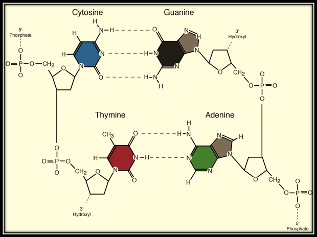
![The image is a structural representation of a DNA double helix. It illustrates the arrangement of nucleotide pairs across the DNA strands.
**Description:**
1. **DNA Structure:**
- The diagram displays two long strands running vertically and intertwined, which compose the DNA backbone. They are connected by pairs of nitrogenous bases.
- The backbone consists of sugar and phosphate groups.
2. **Nitrogenous Bases:**
- The bases are connected via hydrogen bonds, shown by dotted lines between them.
- Each base pairing consists of a purine (adenine [A] or guanine [G]) pairing with a pyrimidine (thymine [T] or cytosine [C]) on the opposite strand.
- The standard base pairs in DNA are adenine with thymine (A-T) and guanine with cytosine (G-C).
3. **Orientation:**
- The top and bottom of the diagram are labeled “TOP” and “BOTTOM” indicating the 5' to 3' orientation of the DNA strands.
4. **Hydrogen Bonds:**
- Hydrogen bonds are depicted as dashed lines between the base pairs, illustrating the specific pairing (A with T, G with C).
5. **Overall Structure:**
- The pattern repeats itself along the strands, indicating the repeating, alternating nature of the bases within the DNA molecule.
This diagram emphasizes the structural organization of DNA, crucial for its role in genetic information storage and transmission.](/v2/_next/image?url=https%3A%2F%2Fcontent.bartleby.com%2Fqna-images%2Fquestion%2Fadc447ea-4c16-4666-abc4-68a3d9e32223%2F12e659c7-5d59-4c90-8745-a0eca4993ff5%2F76z3nc_processed.png&w=3840&q=75)
DNA or deoxyribonucleic acid is the genetic material present in most organisms. It is composed of four nucleotide bases, adenine, guanine, thymine, and cytosine. It is a double helical structure where the strands are present in the opposite polarity of each other. One strand runs from 5' to 3' while the other runs from 3' to 5'
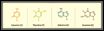
The Adenine and thymine residue joins by two hydrogen bonds, while there are three hydrogen bonds between the guanine and cytosine residue.

Step by step
Solved in 2 steps with 2 images


