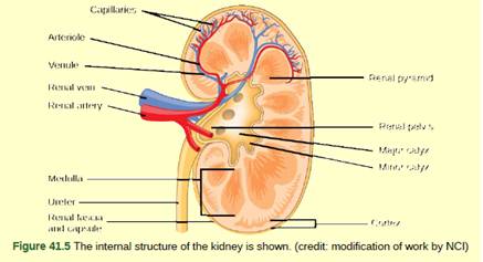
Figure 41.5 Which of the following statements about the kidney is false?
- The renal pelvis drains into the ureter.

Introduction:
In the body, kidneys are found as the pair of bear-shaped structure which are found below the liver in the peritoneal cavity. These bots structures play the functions of the major excretory system as it undergoes the ultrafiltration of the blood.
Answer to Problem 1VCQ
Correct answer:
The correct answer is option (c) The cortex covers the capsule.
Explanation of Solution
Explanation/justification for the correct answer:
Option(c) The cortex covers the capsule. Renal cortex is found as the outer region of the kidney which is formed by the outer extensions of cortical tissue. It is found as the outer portion present between the renal capsule and renal medulla. So it does not found as the covering of capsule only.
Explanation for incorrect answer:
Option (a). Renal pelvis drains into the ureter. The renal pelvis is found as the inner region of the kidney that enters into the ureter which is present outside the kidney. So, this is an incorrect option.
Option (b) Renal pyramids are in the medulla. Renal Medulla is found at the medial region of the kidney which contains many renal pyramids. These pyramids are found as the pyramidal masses of tissues. So, this is an incorrect answer.
Option (d) Nephrons are in the renal cortex. The renal cortex contains the millions of structure and functional unit of the kidney known as nephrons.
Renal cortex is found as the outer region other of the kidney which is formed by the outer extensions of cortical tissue. Hence, the correct answer in s an option (c) The cortex covers the capsule.
Want to see more full solutions like this?
Chapter 41 Solutions
Biology 2e
Additional Science Textbook Solutions
Biological Science (6th Edition)
Campbell Biology in Focus (2nd Edition)
Concepts of Genetics (12th Edition)
Biology: Life on Earth (11th Edition)
College Physics: A Strategic Approach (3rd Edition)
Anatomy & Physiology (6th Edition)
- Use the following tree diagram to answer Questions #8-10. 8) Which of the following two animals are the most closely related based on the tree to the left? a) Pig and camel b) Hippo and pig c) Deer and cow 9) CIRCLE on the tree diagram where the common ancestor between a hippo and a cow is. 10) Put a SQUARE on the tree diagram where the common ancestor between a pig and a peccary is.arrow_forwardExplain: Healthy Cell Function Overview→ Briefly describe how a healthy cell usually works: metabolism (ATP production), pH balance, glycogen storage, ion transport, enzymes, etc. Gene Mutation and Genetics Part→ Focus on the autosomal recessive mutation and explain: How gene mutation affects the cell. How autosomal inheritance works. Compare the normal and mutated gene sequences simply. → Talk about possible consequences of a faulty hydrolytic enzyme.arrow_forwardCan you fill out those termsarrow_forward
- Explain down bellow what happens to the cell: Decreased pH in mitochondria Increased ATP Decreased pH in cytosol Increased hydrolysis Decreasing glycogen and triglycerides Increased MAP kinase activity Poor ion transport → For each one:→ What normally happens?→ What is wrong now?→ How does it mess up the cell?arrow_forwardAn 1100 pound equine patient was given 20 mg/kg sucralfate 3 times a day, 2.8 mg/kg famotidine twice a day, and 10mg/kg doxycycline twice a day. Sucralfate comes as a 1 gm tablet, famotidine as 20 mg tablets, and doxycycline as 100mg tablets. All are in bottles of 100 tablets.How many total mg are needed for the patient and how many tablets of each would be needed to provide each dose?How many bottles of each would be needed to have available if this patient were to be on this drug regimen for 5 days?arrow_forwardThe patient needs a solution of 2.5% dextrose in Lactated Ringer’s solution to run at 75 ml/hr for at least the next 12hours. LRS comes in fluid bags of 500 ml, 1 Liter, 3 Liters and 5 Liters. How can a 2.5% solution be made by adding50% dextrose to the LRS?arrow_forward
- “Gretchen” was a 68-pound canine who came to the VMTH as small animal surgery patient. She receivedacepromazine, 0.2 mg/kg from a 10 mg/ml solution and oxymorphone, 0.08 mg/kg from a 1 mg/ml solution before surgery.What are the mechanisms of action of acepromazine and oxymorphone? Why would they be given together?How many mg provide each dose and how many ml of each of these solutions were given?arrow_forwardAfter surgery, “Gretchen” was put on carprofen, 1 mg/pound bid (twice a day). The tablets come in 25, 75 and 100 mgsizes. Which size tablet would be appropriate?What is the mechanism of action of carprofen?An outpatient prescription was written for her so she would have enough for 10 days. How many tablets did she need?What information needs to be on her out-patient prescription?arrow_forwardJoden Koepp olor in chickens is due to incomplete dominance. BB = Black chicken, WW = White BLOOD TYPES Arhite chicken is In humans, Rh positive blood is dominant (R) over Rh negative blood (r). A man with type 0, Rh positive blood (whose mother had Rh negative blood), marries a woman with type AB, Rh negative blood. Several children were born. is? R R Genotypes Phenotypes RRR RR Rr Rr 4/16 RR R RR RK Rr Rr 4/16 rr 3/4 Rh posi 1/4 Rh negu 1/2 Rr rr rr rrrr 88 888 75 e genotype of the man? the genotype of the woman? The mother of the man had type AB blood.arrow_forward
 Biology 2eBiologyISBN:9781947172517Author:Matthew Douglas, Jung Choi, Mary Ann ClarkPublisher:OpenStax
 Biology (MindTap Course List)BiologyISBN:9781337392938Author:Eldra Solomon, Charles Martin, Diana W. Martin, Linda R. BergPublisher:Cengage Learning


