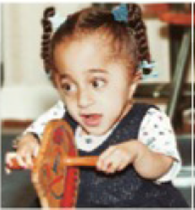
Building Better Bones Tiffany, shown in FIGURE 35.22, was born with multiple fractures in her arms and legs. By age six, she had undergone surgery to correct more than 200 bone fractures. Her fragile, easily broken bones are symptoms of osteogenesis imperfecta (OI), a genetic disorder caused by a mutation in a gene for collagen. As bones develop, collagen forms a scaffold for deposition of mineralized bone tissue. The scaffold forms improperly in children with OI. FIGURE 35.22 also shows the results of a test of a new drug. Treated children, all less than two years old, were compared to similarly affected children of the same age who were not treated with the drug.
| Vertebral | |||
| Treated | area in cm2 | Fractures | |
| child | (Initial) | (Final) | per year |
| 1 | 14.7 | 16.7 | 1 |
| 2 | 15.5 | 16.9 | 1 |
| 3 | 6.7 | 16.5 | 6 |
| 4 | 7.3 | 11.8 | 0 |
| 5 | 13.6 | 14.6 | 6 |
| 6 | 9.3 | 15.6 | 1 |
| 7 | 15.3 | 15.9 | 0 |
| 8 | 9.9 | 13.0 | 4 |
| 9 | 10.5 | 13.4 | 4 |
| Mean | 11.4 | 14.9 | 2.6 |
| Vertebral | |||
| Treated | area in cm2 | Fractures | |
| child | (Initial) | (Final) | per year |
| 1 | 18.2 | 13.7 | 4 |
| 2 | 16.5 | 12.9 | 7 |
| 3 | 16.4 | 11.3 | 8 |
| 4 | 13.5 | 7.7 | 5 |
| 5 | 16.2 | 16.1 | 8 |
| 6 | 18.9 | 17.0 | 6 |
| Mean | 16.6 | 13.1 | 6.3 |

FIGURE 35.22 Results of a clinical trial of a drug treatment for osteogenesis imperfecta (OI), which affects the child shown at right. Nine children with OI received the drug. Six others were untreated controls. Surface area of certain vertebrae was measured before and after treatment. Fractures occurring during the 12 months of the trial were also recorded.
3. How did the rate of fractures in the two groups compare?
Want to see the full answer?
Check out a sample textbook solution
Chapter 35 Solutions
Bundle: Biology: The Unity and Diversity of Life, Loose-leaf Version, 14th + LMS Integrated for MindTap Biology, 2 terms (12 months) Printed Access Card
- What happens to a microbes membrane at colder temperature?arrow_forwardGenes at loci f, m, and w are linked, but their order is unknown. The F1 heterozygotes from a cross of FFMMWW x ffmmww are test crossed. The most frequent phenotypes in the test cross progeny will be FMW and fmw regardless of what the gene order turns out to be. What classes of testcross progeny (phenotypes) would be least frequent if locus m is in the middle? What classes would be least frequent if locus f is in the middle? What classes would be least frequent if locus w is in the middle?arrow_forward1. In the following illustration of a phospholipid... (Chemistry Primer and Video 2-2, 2-3 and 2-5) a. Label which chains contain saturated fatty acids and non-saturated fatty acids. b. Label all the areas where the following bonds could form with other molecules which are not shown. i. Hydrogen bonds ii. Ionic Bonds iii. Hydrophobic Interactions 12-6 HICIH HICIH HICHH HICHH HICIH OHHHHHHHHHHHHHHHHH C-C-C-C-C-c-c-c-c-c-c-c-c-c-c-c-C-C-H HH H H H H H H H H H H H H H H H H H HO H-C-O H-C-O- O O-P-O-C-H H T HICIH HICIH HICIH HICIH HHHHHHH HICIH HICIH HICIH 0=C HIC -C-C-C-C-C-C-C-C-CC-C-C-C-C-C-C-C-C-H HHHHHHHHH IIIIIIII HHHHHHHH (e-osbiv)arrow_forward
- O Macmillan Learning Glu-His-Trp-Ser-Gly-Leu-Arg-Pro-Gly The pKa values for the peptide's side chains, terminal amino groups, and carboxyl groups are provided in the table. Amino acid Amino pKa Carboxyl pKa Side-chain pKa glutamate 9.60 2.34 histidine 9.17 1.82 4.25 6.00 tryptophan 9.39 2.38 serine 9.15 2.21 glycine 9.60 2.34 leucine 9.60 2.36 arginine 9.04 2.17 12.48 proline 10.96 1.99 Calculate the net charge of the molecule at pH 3. net charge at pH 3: Calculate the net charge of the molecule at pH 8. net charge at pH 8: Calculate the net charge of the molecule at pH 11. net charge at pH 11: Estimate the isoelectric point (pl) for this peptide. pl:arrow_forwardBiology Questionarrow_forwardThis entire structure (Pinus pollen cone) using lifecycle terminology is called what?arrow_forward
- This entire structure using lifecycle terminology is called what? megastrobilus microstrobilus megasporophyll microsporophyll microsporangium megasporangium none of thesearrow_forwardHow much protein should Sarah add to her diet if she gets pregnant? Sarah's protein requirements during pregnancy would be higher. See Hint B2. During calculations, use numbers rounded to the first decimal place. In your answer, round the number of grams to the nearest whole number. _______ g ?arrow_forwardC MasteringHealth MasteringNu X session.healthandnutrition-mastering.pearson.com/myct/itemView?assignment ProblemID=17396422&attemptNo=1&offset=prevarrow_forwardarrow_back_iosSEE MORE QUESTIONSarrow_forward_ios
 Biology: The Dynamic Science (MindTap Course List)BiologyISBN:9781305389892Author:Peter J. Russell, Paul E. Hertz, Beverly McMillanPublisher:Cengage Learning
 Anatomy & PhysiologyBiologyISBN:9781938168130Author:Kelly A. Young, James A. Wise, Peter DeSaix, Dean H. Kruse, Brandon Poe, Eddie Johnson, Jody E. Johnson, Oksana Korol, J. Gordon Betts, Mark WomblePublisher:OpenStax College
 Biology: The Unity and Diversity of Life (MindTap...BiologyISBN:9781305073951Author:Cecie Starr, Ralph Taggart, Christine Evers, Lisa StarrPublisher:Cengage Learning
 Biology 2eBiologyISBN:9781947172517Author:Matthew Douglas, Jung Choi, Mary Ann ClarkPublisher:OpenStax
 Biology: The Unity and Diversity of Life (MindTap...BiologyISBN:9781337408332Author:Cecie Starr, Ralph Taggart, Christine Evers, Lisa StarrPublisher:Cengage Learning
 Principles Of Radiographic Imaging: An Art And A ...Health & NutritionISBN:9781337711067Author:Richard R. Carlton, Arlene M. Adler, Vesna BalacPublisher:Cengage Learning


