
Videos
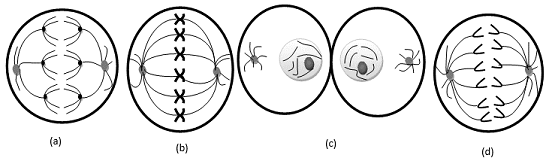
Examine the following diagrams of cells from an organism with diploid number 2n = 6, and identify what stage of M phase is represented.

To review:
Identification of stages of mitotic or meiotic phase from the given diagrams in question.
Introduction:
There are two types of cell division – mitosis and meiosis. Somatic cell division is termed as mitosis, and germ cell division is termed as meiosis. The prophase, metaphase, anaphase, and telophase are four phases in mitosis. In prophase, chromatin gets condensed and chromosomes become visible under microscope. In metaphase, homologous chromosomes get aligned in the center of the cell. In anaphase, sister chromatids are separated by mitotic spindles. The last phase is telophase in which a nuclear membrane is formed between daughter cells; a cleavage furrow is formed which separates the cytoplasm of the parent cell equally, and diploid daughter cells are formed from diploid parent cells.
Meiosis 1 and meiosis 2 are the two cycles in meiosis. Meiosis 1 is known as reduction division. The rare event of crossing over occurs in this phase in which segments of non-sister chromatids are exchanged. Meiosis 1 results in two haploid daughter cells, and then meiosis 2 begins, which forms 4 haploid cells.
Explanation of Solution
The given diagram is of anaphase 1. The given diagram shows that the homologous chromosomes get pulled by microtubules and move to opposite poles. This phenomenon is observed in both types of cell division (mitosis and meiosis), but here, segments of chromosomes are shown in different colors, which means crossing over takes place between them, and crossing over is s rare phenomenon, which occurs in meiosis 1 (Anaphase 1) only.
The given diagram is of metaphase. It can be concluded by observing the position of chromosomes. In the given diagram, chromosomes are aligned in the middle of the cell.
The given diagram is of cytokinesis (telophase). Cleavage furrow is formed between daughter cells which separates parent cytoplasm equally into two daughter cells. Initiation of nuclear membrane formation is observed.
Given diagram is of anaphase. Initiation of movement of sister chromatids to apposite pole is observed as sister chromatids get pulled by mitotic spindles. Crossing over is not observed.
The given diagram is of anaphase 1.
The given diagram is of metaphase.
The given diagram is of cytokinesis (telophase).
The given diagram is of anaphase.
Want to see more full solutions like this?
Chapter 3 Solutions
Genetic Analysis: An Integrated Approach (3rd Edition)
Additional Science Textbook Solutions
Biology: Life on Earth with Physiology (11th Edition)
Chemistry: The Central Science (14th Edition)
Chemistry: Structure and Properties (2nd Edition)
Applications and Investigations in Earth Science (9th Edition)
Physics for Scientists and Engineers: A Strategic Approach, Vol. 1 (Chs 1-21) (4th Edition)
Campbell Biology in Focus (2nd Edition)


- State the five functions of Globular Proteins, and give an example of a protein for each function.arrow_forwardDiagram of check cell under low power and high powerarrow_forwarda couple in which the father has the a blood type and the mother has the o blood type produce an offspring with the o blood type, how does this happen? how could two functionally O parents produce an offspring that has the a blood type?arrow_forward
- What is the opening indicated by the pointer? (leaf x.s.) stomate guard cell lenticel intercellular space none of thesearrow_forwardIdentify the indicated tissue? (stem x.s.) parenchyma collenchyma sclerenchyma ○ xylem ○ phloem none of thesearrow_forwardWhere did this structure originate from? (Salix branch root) epidermis cortex endodermis pericycle vascular cylinderarrow_forward
- Identify the indicated tissue. (Tilia stem x.s.) parenchyma collenchyma sclerenchyma xylem phloem none of thesearrow_forwardIdentify the indicated structure. (Cucurbita stem l.s.) pit lenticel stomate tendril none of thesearrow_forwardIdentify the specific cell? (Zebrina leaf peel) vessel element sieve element companion cell tracheid guard cell subsidiary cell none of thesearrow_forward
- What type of cells flank the opening on either side? (leaf x.s.) vessel elements sieve elements companion cells tracheids guard cells none of thesearrow_forwardWhat specific cell is indicated. (Cucurbita stem I.s.) vessel element sieve element O companion cell tracheid guard cell none of thesearrow_forwardWhat specific cell is indicated? (Aristolochia stem x.s.) vessel element sieve element ○ companion cell O O O O O tracheid O guard cell none of thesearrow_forward
 Human Heredity: Principles and Issues (MindTap Co...BiologyISBN:9781305251052Author:Michael CummingsPublisher:Cengage Learning
 Human Biology (MindTap Course List)BiologyISBN:9781305112100Author:Cecie Starr, Beverly McMillanPublisher:Cengage Learning
 Biology (MindTap Course List)BiologyISBN:9781337392938Author:Eldra Solomon, Charles Martin, Diana W. Martin, Linda R. BergPublisher:Cengage Learning
 Concepts of BiologyBiologyISBN:9781938168116Author:Samantha Fowler, Rebecca Roush, James WisePublisher:OpenStax College
 Biology 2eBiologyISBN:9781947172517Author:Matthew Douglas, Jung Choi, Mary Ann ClarkPublisher:OpenStax