
Videos
To review:
The sequence of events that occurs during the cardiac cycle through flow chart and the indication of contraction of auricle and ventricle and their relaxation.
Introduction:
Circulatory system circulates the blood into two directions simultaneously, one from the heart to the body known as systematic circulation and the other from the body to the heart, from where it goes to the lungs for purification called the pulmonary circulation and the whole process is known as double circulation.
Explanation:
The right side of the heart contains blood which is poor in oxygen and the left side contains oxygen-rich blood, the atrioventricular (AV) valve and semilunar valves that are present between the auricle and ventricle on their side and at the opening of pulmonary artery and aorta, respectively. These prevent the back flow in the heart. The right auricle is usually filled with the deoxygenated blood by superior or inferior vena cava and the left auricle is filled by the oxygenated blood coming from lungs by the pulmonary veins.
The contraction (systole) of auricles on both sides start after they are completely filled with the blood and the blood enters in their corresponding ventricles through AV valves, as soon as the ventricles are filled up and starts contracting. The AV valves are closed to prevent the back flow of blood and starts auricular relaxation. The blood from right ventricle moves to the lungs for its purification through pulmonary artery and oxygenated blood from left ventricle is transported to other body parts through the aorta. This completes one cycle of double circulation of blood in the body and it is known as a cardiac cycle, and this cycle is continuous. The diagram below shows the steps of cardiac cycle:
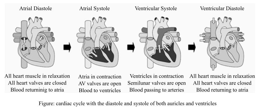
Diastole means relaxation of the auricles and ventricles and systole is the contraction of auricles and ventricles. Atrial diastole occurs when the blood returns to the atria from vena cava on right side and from pulmonary vein on left side. The valves of the heart, during this time, are closed and muscles are relaxed. When the atria are filled, the AV valves open and atrial systole occurs. The ventricular systole occurs when they are completely filled and semilunar valves open and blood releases out from the ventricles and cause diastole of ventricles. This way the cardiac cycle goes on till the heart is functional.
Explanation:
The right side of the heart contains blood which is poor in oxygen and the left side contains oxygen-rich blood, the atrioventricular (AV) valve and semilunar valves that are present between the auricle and ventricle on their side and at the opening of pulmonary artery and aorta, respectively. These prevent the back flow in the heart. The right auricle is usually filled with the deoxygenated blood by superior or inferior vena cava and the left auricle is filled by the oxygenated blood coming from lungs by the pulmonary veins.
The contraction (systole) of auricles on both sides start after they are completely filled with the blood and the blood enters in their corresponding ventricles through AV valves, as soon as the ventricles are filled up and starts contracting. The AV valves are closed to prevent the back flow of blood and starts auricular relaxation. The blood from right ventricle moves to the lungs for its purification through pulmonary artery and oxygenated blood from left ventricle is transported to other body parts through the aorta. This completes one cycle of double circulation of blood in the body and it is known as a cardiac cycle, and this cycle is continuous. The diagram below shows the steps of cardiac cycle:
Diastole means relaxation of the auricles and ventricles and systole is the contraction of auricles and ventricles. Atrial diastole occurs when the blood returns to the atria from vena cava on right side and from pulmonary vein on left side. The valves of the heart, during this time, are closed and muscles are relaxed. When the atria are filled, the AV valves open and atrial systole occurs. The ventricular systole occurs when they are completely filled and semilunar valves open and blood releases out from the ventricles and cause diastole of ventricles. This way the cardiac cycle goes on till the heart is functional.
Want to see the full answer?
Check out a sample textbook solution
Chapter 13 Solutions
Human Physiology


- Please indentify the unknown organismarrow_forward5G JA ATTC 3 3 CTIA A1G5 5 GAAT I I3 3 CTIA AA5 Fig. 5-3: The Eco restriction site (left) would be cleaved at the locations indicated by the arrows. However, a SNP in the position shown in gray (right) would prevent cleavage at this site by EcoRI One of the SNPs in B. rapa is found within the Park14 locus and can be detected by RFLP analysis. The CT polymorphism is found in the intron of the Bra013780 gene found on Chromosome 1. The Park14 allele with the "C" in the SNP has two EcoRI sites and thus is cleaved twice by EcoRI If there is a "T" in that SNP, one of the EcoRI sites is altered, so the Park14 allele with the T in the SNP has only one EcoRI site (Fig. 5-3). Park14 allele with SNP(C) Park14 allele with SNPT) 839 EcoRI 1101 EcoRI 839 EcoRI Fig. 5.4: Schematic restriction maps of the two different Park14 alleles (1316 bp long) of B. rapa. Where on these maps is the CT SNP located? 90 The primers used to amplify the DNA at the Park14 locus (see Fig. 5 and Table 3 of Slankster et…arrow_forwardFrom your previous experiment, you found that this enhancer activates stripe 2 of eve expression. When you sequence this enhancer you find several binding sites for the gap gene, Giant. To test how Giant interacts with eve, you decide to remove all of the Giant binding sites from the eve enhancer. What results do you expect to see with respect to eve expression?arrow_forward
- What experiment could you do to see if the maternal gene, bicoid, is sufficient to form anterior fates?arrow_forwardYou’re curious about the effect that gap genes have on the pair-rule gene, evenskipped (eve), so you isolate and sequence each of the eve enhancers. You’re particularly interested in one of the enhancers, which is just upstream of the eve gene. Describe an experimental technique you would use to find out where this particular eve enhancer is active.arrow_forwardFor short answer questions, write your answers on the line provided. To the right is the mRNA codon table to use as needed throughout the exam. First letter U บบบ U CA UUCPhe UUA UCU Phe UCC UUG Leu CUU UAU. G U UAC TV UGCys UAA Stop UGA Stop A UAG Stop UGG Trp Ser UCA UCG CCU] 0 CUC CUA CCC CAC CAU His CGU CGC Leu Pro CCA CAA Gin CGA Arg CUG CCG CAG CGG AUU ACU AAU T AUC lle A 1 ACC Thr AUA ACA AUG Mot ACG AGG Arg GUU GCU GUC GCC G Val Ala GAC Asp GGU GGC GUA GUG GCA GCG GAA GGA Gly Glu GAGJ GGG AACASH AGU Ser AAA1 AAG Lys GAU AGA CAL CALUCAO CAO G Third letter 1. (+7) Use the table below to answer the questions; use the codon table above to assist you. The promoter sequence of DNA is on the LEFT. You do not need to fill in the entire table. Assume we are in the middle of a gene sequence (no need to find a start codon). DNA 1 DNA 2 mRNA tRNA Polypeptide C Val G C. T A C a. On which strand of DNA is the template strand (DNA 1 or 2)?_ b. On which side of the mRNA is the 5' end (left or…arrow_forward
- 3. (6 pts) Fill in the boxes according to the directions on the right. Structure R-C R-COOH OH R-OH i R-CO-R' R R-PO4 R-CH3 C. 0 R' R-O-P-OH 1 OH H R-C-H R-N' I- H H R-NH₂ \H Name Propertiesarrow_forward4. (6 pts) Use the molecule below to answer these questions and identify the side chains and ends. Please use tidy boxes to indicate parts and write the letter labels within that box. a. How many monomer subunits are shown? b. Box a Polar but non-ionizable side chain and label P c. Box a Basic Polar side chain and label BP d. Box the carboxyl group at the end of the polypeptide and label with letter C (C-terminus) H H OHHO H H 0 HHO H-N-CC-N-C-C N-C-C-N-GC-OH I H-C-H CH2 CH2 CH2 H3C-C+H CH2 CH2 OH CH CH₂ C=O OH CH2 NH2arrow_forwardplease answer (A,B,C,D,E) questions with the asnwer choice provided below. thank you!arrow_forward
 Human Physiology: From Cells to Systems (MindTap ...BiologyISBN:9781285866932Author:Lauralee SherwoodPublisher:Cengage Learning
 Human Biology (MindTap Course List)BiologyISBN:9781305112100Author:Cecie Starr, Beverly McMillanPublisher:Cengage Learning
- Basic Clinical Lab Competencies for Respiratory C...NursingISBN:9781285244662Author:WhitePublisher:Cengage
 Fundamentals of Sectional Anatomy: An Imaging App...BiologyISBN:9781133960867Author:Denise L. LazoPublisher:Cengage Learning