
Videos
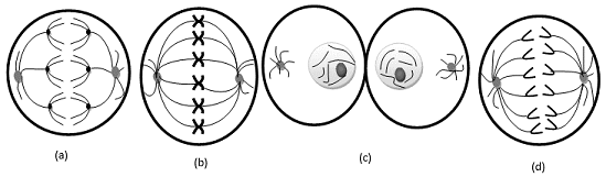
Examine the following diagrams of cells from an organism with diploid number 2n = 6, and identify what stage of M phase is represented.

To review:
Identification of stages of mitotic or meiotic phase from the given diagrams in question.
Introduction:
There are two types of cell division – mitosis and meiosis. Somatic cell division is termed as mitosis, and germ cell division is termed as meiosis. The prophase, metaphase, anaphase, and telophase are four phases in mitosis. In prophase, chromatin gets condensed and chromosomes become visible under microscope. In metaphase, homologous chromosomes get aligned in the center of the cell. In anaphase, sister chromatids are separated by mitotic spindles. The last phase is telophase in which a nuclear membrane is formed between daughter cells; a cleavage furrow is formed which separates the cytoplasm of the parent cell equally, and diploid daughter cells are formed from diploid parent cells.
Meiosis 1 and meiosis 2 are the two cycles in meiosis. Meiosis 1 is known as reduction division. The rare event of crossing over occurs in this phase in which segments of non-sister chromatids are exchanged. Meiosis 1 results in two haploid daughter cells, and then meiosis 2 begins, which forms 4 haploid cells.
Explanation of Solution
The given diagram is of anaphase 1. The given diagram shows that the homologous chromosomes get pulled by microtubules and move to opposite poles. This phenomenon is observed in both types of cell division (mitosis and meiosis), but here, segments of chromosomes are shown in different colors, which means crossing over takes place between them, and crossing over is s rare phenomenon, which occurs in meiosis 1 (Anaphase 1) only.
The given diagram is of metaphase. It can be concluded by observing the position of chromosomes. In the given diagram, chromosomes are aligned in the middle of the cell.
The given diagram is of cytokinesis (telophase). Cleavage furrow is formed between daughter cells which separates parent cytoplasm equally into two daughter cells. Initiation of nuclear membrane formation is observed.
Given diagram is of anaphase. Initiation of movement of sister chromatids to apposite pole is observed as sister chromatids get pulled by mitotic spindles. Crossing over is not observed.
The given diagram is of anaphase 1.
The given diagram is of metaphase.
The given diagram is of cytokinesis (telophase).
The given diagram is of anaphase.
Want to see more full solutions like this?
Chapter 3 Solutions
Genetic Analysis: An Integrated Approach (3rd Edition)
Additional Science Textbook Solutions
Biology: Life on Earth with Physiology (11th Edition)
Chemistry: The Central Science (14th Edition)
Chemistry: Structure and Properties (2nd Edition)
Applications and Investigations in Earth Science (9th Edition)
Physics for Scientists and Engineers: A Strategic Approach, Vol. 1 (Chs 1-21) (4th Edition)
Campbell Biology in Focus (2nd Edition)


- What is this?arrow_forwardMolecular Biology A-C components of the question are corresponding to attached image labeled 1. D component of the question is corresponding to attached image labeled 2. For a eukaryotic mRNA, the sequences is as follows where AUGrepresents the start codon, the yellow is the Kozak sequence and (XXX) just represents any codonfor an amino acid (no stop codons here). G-cap and polyA tail are not shown A. How long is the peptide produced?B. What is the function (a sentence) of the UAA highlighted in blue?C. If the sequence highlighted in blue were changed from UAA to UAG, how would that affecttranslation? D. (1) The sequence highlighted in yellow above is moved to a new position indicated below. Howwould that affect translation? (2) How long would be the protein produced from this new mRNA? Thank youarrow_forwardMolecular Biology Question Explain why the cell doesn’t need 61 tRNAs (one for each codon). Please help. Thank youarrow_forward
- Molecular Biology You discover a disease causing mutation (indicated by the arrow) that alters splicing of its mRNA. This mutation (a base substitution in the splicing sequence) eliminates a 3’ splice site resulting in the inclusion of the second intron (I2) in the final mRNA. We are going to pretend that this intron is short having only 15 nucleotides (most introns are much longer so this is just to make things simple) with the following sequence shown below in bold. The ( ) indicate the reading frames in the exons; the included intron 2 sequences are in bold. A. Would you expected this change to be harmful? ExplainB. If you were to do gene therapy to fix this problem, briefly explain what type of gene therapy youwould use to correct this. Please help. Thank youarrow_forwardMolecular Biology Question Please help. Thank you Explain what is meant by the term “defective virus.” Explain how a defective virus is able to replicate.arrow_forwardMolecular Biology Explain why changing the codon GGG to GGA should not be harmful. Please help . Thank youarrow_forward
- Stage Percent Time in Hours Interphase .60 14.4 Prophase .20 4.8 Metaphase .10 2.4 Anaphase .06 1.44 Telophase .03 .72 Cytukinesis .01 .24 Can you summarize the results in the chart and explain which phases are faster and why the slower ones are slow?arrow_forwardCan you circle a cell in the different stages of mitosis? 1.prophase 2.metaphase 3.anaphase 4.telophase 5.cytokinesisarrow_forwardWhich microbe does not live part of its lifecycle outside humans? A. Toxoplasma gondii B. Cytomegalovirus C. Francisella tularensis D. Plasmodium falciparum explain your answer thoroughly.arrow_forward
- Select all of the following that the ablation (knockout) or ectopoic expression (gain of function) of Hox can contribute to. Another set of wings in the fruit fly, duplication of fingernails, ectopic ears in mice, excess feathers in duck/quail chimeras, and homeosis of segment 2 to jaw in Hox2a mutantsarrow_forwardSelect all of the following that changes in the MC1R gene can lead to: Changes in spots/stripes in lizards, changes in coat coloration in mice, ectopic ear formation in Siberian hamsters, and red hair in humansarrow_forwardPleiotropic genes are genes that (blank) Cause a swapping of organs/structures, are the result of duplicated sets of chromosomes, never produce protein products, and have more than one purpose/functionarrow_forward
 Human Heredity: Principles and Issues (MindTap Co...BiologyISBN:9781305251052Author:Michael CummingsPublisher:Cengage Learning
 Human Biology (MindTap Course List)BiologyISBN:9781305112100Author:Cecie Starr, Beverly McMillanPublisher:Cengage Learning
 Biology (MindTap Course List)BiologyISBN:9781337392938Author:Eldra Solomon, Charles Martin, Diana W. Martin, Linda R. BergPublisher:Cengage Learning
 Concepts of BiologyBiologyISBN:9781938168116Author:Samantha Fowler, Rebecca Roush, James WisePublisher:OpenStax College
 Biology 2eBiologyISBN:9781947172517Author:Matthew Douglas, Jung Choi, Mary Ann ClarkPublisher:OpenStax