Videos
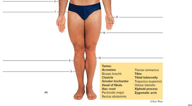
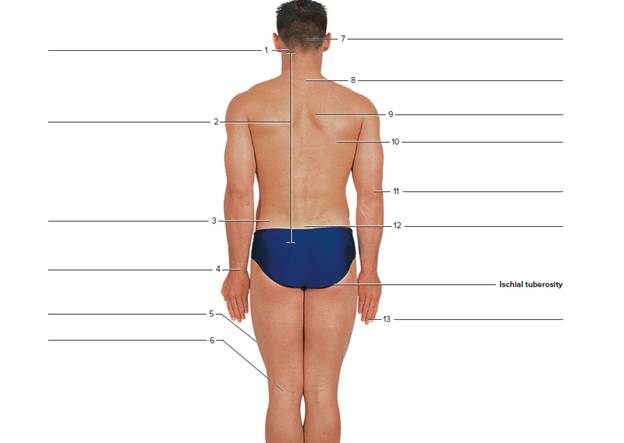
FIGURE 26.9 Using the terms provided, label the surface features of the (a) anterior view of the body and (b) posterior view of the body (Boldface indicates bony features: not boldface indicates soft tissue features.)


Terms:
C7 					lliac crest 			Olecranon process
Calcaneal tendon 			lliotibiai tract 		PopiteaI fossa
Distal Interphalangeal Joint (DIP) 	Inferior angle of scapula 	Sacrum
Erector spinae				Mastoid process 		Soleus
External occipItal protuberance 	Medial border of scapula 	Stylold process of ulna
To label:
The given image in anterior and posterior view of the human body.
Introduction:
Skeleton is a rigid bony or cartilaginous protective framework in an organism. It generally protects the soft tissues and internal organs of a vertebrate.The human skeleton plays six main roles that include the movement, protection, support, storage of minerals, production of blood cells, and endocrine regulation.
Explanation of Solution
Muscle is a bundle or band of fibrous tissue present in a human or animal body. It can contract and move in preserving the position of body parts. Muscles play a role in the production of force and motion. They are mainly responsible for sustaining and shifting posture, locomotion.
The anterior view:
1. Zygomatic arch:
Zygomatic arch is a bridge of bone that extends from the temporal bone at around the head side to the upper jawbone in front and has the cheek or zygomatic bone as its main region.
2. Trapezius:
A large paired surface muscle is the trapezius. It spreads longitudinally from the region of occipital bone to the area of lower thoracic vertebrae and across the spine of the scapula. It is a flat, broad, and superficial muscle.
It is responsible for rotating, moving, and stabilizing the shoulder blade or scapula.
3. Clavicle:
The pectoral girdle or shoulder girdle is a set of bones located in the appendicular skeleton. It joins to the arm on each side. Pectoral girdle consists of the scapula, coracoid and clavicle.
Clavicle is a bone of the pectoral girdle usually helps to join the scapula and sternum is known as the collarbone.
4. Xiphoid process:
It is a cartilaginous section located at the inferior end of the sternum. It is not associated to any ribs, steadily ossifies through the adult life.
5. Rectus abdominis:
It is also known as abdominal muscle. It is a paired muscle. It runs vertically on both the sides of the anterior wall of the abdomen (in human) in addition to some other mammals. It flexes trunk of the body or the spinal column.
6. Iliac crest: It is a curved superior boundary of the ilium. It is the biggest of the three bones. It merges to shape the hip bone or os coxa.
7. Greater trochanter:
A trochanter is a type of tubercle of the femur. It is close to the joint with the hip bone. These trochanters play a great role as a muscle attachment site in most of the mammals and humans. Humans have three trochanters. Though the normal anatomic has only the greater and lesser trochanters. A trochanter is a very huge projection on a bone.
8. Tibial tuberosity:
Tuberosity is a moderate distinction where the connective tissues and muscles join. Its role is similar to the trochanter. For example the tibial tuberosity. The tuberosity of the tibia or tibial tuberosity or tibial tubercle is a large oblong elevation on the proximal, anterior aspect of the tibia, just below where the anterior surfaces of the lateral and medial tibial condyles end.
9. Tibia:
It is the inner and characteristically larger of the two bones located between the knee and the ankle. It is parallel with the fibula.
10. Acromion:
It is a bony process located on the scapula or shoulder blade. It expands laterally over the shoulder joint along with the coracoid process. It is an extension of the scapular spine that hooks over anteriorly.
11. Pectoralis major:
The pectoralis major is a thick and fan-shaped muscle. It is situated at the human body chest. It forms up the bulkiness of the muscles of the chest. It is located under the breast.
12. The biceps:
The brachialis anticus or the brachialis is a muscle located in the upper arm. It flexes the elbow joint. It is located deeper than the biceps brachii. It makes up the region of the floor of the location that is known as the cubital fossa. The brachialis functions as the major mover of elbow flexion. The flexor group includes the biceps brachii, brachialis, and the brachioradialis. It helps in the bending of the arm by declining the angle present between the upper arm and the forearm.
13. Thenar eminence:
It is classified as the group of muscles located on the human hand palm. It is present at the thumb base.
14. Vastus lateralis:
The vastus lateralis is also known as the vastus externus. It is the largest and powerful region of the quadriceps femoris. It is a muscle of the thigh or lower limb. Along with the other muscles of quadriceps group. It helps to enlarge the knee joint and moves the lower leg forward.
15. Head of fibula:
The fibula (or calf bone) is a leg bone found on the lateral side of the tibia. It is joined above and below to tibia. It is the smaller than tibia in proportion to its length. Fibula is also the slenderest of all the long bones. The head region is the uppermost region of fibula.
The posterior view:
1. Mastoid process:
It is a conical eminence of the temporal bone. It is present behind the ear. It is attached with the help of neck muscles. It contains air spaces connected to the middle ear.
2. Medial border of scapula:
The shoulder blade or scapula or wing bone is the well-known flat triangular bone located at the back of the shoulder. The pectoral girdle or shoulder girdle is a set of bones located in the appendicular skeleton. It joins to the arm on each side. Pectoral girdle consists of the scapula, coracoid and clavicle. It has a medial border.
3. Iliac crest: It is a curved superior boundary of the ilium. It is the biggest of the three bones. It merges to shape the hip bone or os coxa.
4. Styloid process of ulna:
The ulna bone is a long bone located in the forearm. It stretches from the region of elbow to the smallest finger. In a normal anatomical location it lies on the medial side of the forearm. It extends parallel to the radius (the other long bone of the forearm). Thus, ulna is a bone located in the upper limb. At the distal end of the forearm, the styloid process of the ulna is present. This process projects from the medial portion and back portion of the bone;
5. Iliotibial tract:
It is a longitudinal fibrous support associated with the fascia lata (deep fascia of the thigh). The function of this tract is to abduct, extend, and laterally turn around the hip.
6. Popliteal fossa:
The popliteal fossa is also known as the kneepit. It is a superficial depression. It is situated at the backside of the knee joint.
7. External occipital protuberance:
The external occipital protuberance is the nearest the middle of the squamous portion of the occipital bone. It has a highest point that is known as the inion.
8. Vertebra prominens (C7):
It has a distinct long and well-known spinous process. It is palpable from the surface of the skin.Its provides the support to the skull and helps in the head movements back and forth. It also possesses the side to side head movements and also protects the spinal cord.
9. Erector spinae:
These are a group of long muscles which arise near the sacrum and expand vertically up to the length of the back.
10. Inferior angle of scapula:
It is formed by the combination of the lateral and medial borders of the scapula. It is a thick and rough surface. Its posterior side surface affords the link to the teres major and often to a small number of latissimus dorsi fibers.
11. Olecranon process:
It is a process of the ulna. It makes the elbow outer bump. It fits into the fossa of the humerus bone during the extension of arm.
12. Sacrum:
It is a triangular bone located in the lower part of the back. It is formed from the fused vertebrae. It is located between the two hip bones of the pelvis region.
13. Distal interphalangeal joints (DIP):
The articulations present between the hand or foot phalanges are the distal interphalangeal joints.These are in fact the most common position for osteoarthritis (OA) on the body.
14. Soleus:
It is a strong muscle located in the back portion of the lower leg. It extends from just inferior the knee to the heel. It helps in standing and walking.
15. The calcaneal tendon:
It is also known as an Achilles tendon. It is a hard band of the fibrous tissue. It attaches the calf muscles to the calcaneus or heel bone. The soleus and gastrocnemius muscles combine into a single band of tissue that forms the Achilles tendon at the inferior end of the calf.
Skeleton is a rigid bony or cartilaginous protective framework in an organism. It generally protects the soft tissues and internal organs of a vertebrate.Muscle is a bundle or band of fibrous tissue present in a human or animal body. It can contract and move in preserving the position of body parts.
Want to see more full solutions like this?
Chapter 26 Solutions
Laboratory Manual For Human Anatomy & Physiology
Additional Science Textbook Solutions
HUMAN ANATOMY
Biological Science (6th Edition)
Campbell Biology in Focus (2nd Edition)
Chemistry: A Molecular Approach (4th Edition)
Biology: Life on Earth with Physiology (11th Edition)
Genetics: From Genes to Genomes


- What is a gain of function mutation?arrow_forwardMolecular Biology Question: Please help. Thank you Is Southern hybridization's purpose detecting specific nucleotide sequences? How so?arrow_forwardUse the following information to answer the question(s) below. Martin Wikelski and L. Michael Romero (Body size, performance and fitness in Galápagos marine iguanas, Integrative and Comparative Biology 43 [2003]:376-86) measured the snout-to-vent (anus) length of Galápagos marine iguanas and observed the percent survival of different-sized animals, all of the same age. The graph shows the log snout-vent length (SVL, a measure of overall body size) plotted against the percent survival of these different size classes for males and females. Survival (%) 100- 80- 60- 40- 20- 0+ 1.9 T 2 2.1 2.2 2.3 2.4 2.5 2.6 2.7 Log SVL (mm) 19) Examine the figure above. What type of selection for body size appears to be occurring in these marine iguanas? A) directional selection B) stabilizing selection C) disruptive selection D) You cannot determine the type of selection from the above information. 3arrow_forward
- 24) Use the following information to answer the question below. Researchers studying a small milkweed population note that some plants produce a toxin and other plants do not. They identify the gene responsible for toxin production. The dominant allele (T) codes for an enzyme that makes the toxin, and the recessive allele (t) codes for a nonfunctional enzyme that cannot produce the toxin. Heterozygotes produce an intermediate amount of toxin. The genotypes of all individuals in the population are determined (see table) and used to determine the actual allele frequencies in the population. TT 0.49 Tt 0.42 tt 0.09 Refer to the table above. Is this population in Hardy-Weinberg equilibrium? A) Yes. C) No; there are more homozygotes than expected. B) No; there are more heterozygotes than expected. D) It is impossible to tell.arrow_forward30) A B CDEFG Refer to the accompanying figure. Which of the following forms a monophyletic group? A) A, B, C, and D B) C and D C) D, E, and F D) E, F, and Garrow_forwardMolecular Biology Question. Please help with step solution and explanation. Thank you: The Polymerase Chain Reaction (PCR) reaction consists of three steps denaturation, hybridization, and elongation. Please describe what occurs in the annealing step of the PCR reaction. (I think annealing step is hybridization). What are the other two steps of PCR, and what are their functions? Next, suppose the Tm for the two primers being used are 54C for Primer A and 67C for Primer B. Regarding annealing step temperature, I have the following choices for the temperature used during the annealing step:(a) 43C (b) 49C (c) 62C (d) 73C Which temperature/temperatures should I choose? What is the corresponding correct explanation, and why would I not use the other temperatures? Have a good day!arrow_forward
- Using the data provided on the mean body mass and horn size of 4-year-old male sheep, draw a scatterplot graph to examine how body mass and horn size changed over time.arrow_forwardPlease write a 500-word report about the intake of saturated fat, sodium, alcoholic beverages, or added sugar in America. Choose ONE of these and write about what is recommended by the Dietary Guidelines for Americans (guideline #4) and why Americans exceed the intake of that nutrient. Explain what we could do as a society and/or individuals to reduce our intake of your chosen nutrient.arrow_forwardWrite a 500-word report indicating how you can change the quantity or quality of TWO nutrients where your intake was LOWER than what is recommended by the Dietary Guidelines for Americans and/or the DRIs. Indicate how the lack of the nutrient may affect your health. For full credit, all of the following points must be addressed and elaborated on in more detail for each nutrient: The name of the nutrient At least 2 main functions of the nutrient (example: “Vitamin D regulates calcium levels in the blood and calcification of bones.”) Your percent intake compared to the RDA/DRI (example “I consumed 50% of the RDA for vitamin D”) Indicate why your intake was below the recommendations (example: “I only had one serving of dairy products and that was why I was below the recommendations for vitamin D”) How would you change your dietary pattern to meet the recommendations? – be sure to list specific foods (example: “I would add a yogurt and a glass of milk to each day in order to increase my…arrow_forward
- Why are nutrient absorption and dosage levels important when taking multivitamins and vitamin and mineral supplements?arrow_forwardI'm struggling with this topic and would really appreciate your help. I need to hand-draw a diagram and explain the process of sexual differentiation in humans, including structures, hormones, enzymes, and other details. Could you also make sure to include these terms in the explanation? . Gonads . Wolffian ducts • Müllerian ducts . ⚫ Testes . Testosterone • Anti-Müllerian Hormone (AMH) . Epididymis • Vas deferens ⚫ Seminal vesicles ⚫ 5-alpha reductase ⚫ DHT - Penis . Scrotum . Ovaries • Uterus ⚫ Fallopian tubes - Vagina - Clitoris . Labia Thank you so much for your help!arrow_forwardRequisition Exercise A phlebotomist goes to a patient’s room with the following requisition. Hometown Hospital USA 125 Goodcare Avenue Small Town, USAarrow_forward
 Medical Terminology for Health Professions, Spira...Health & NutritionISBN:9781305634350Author:Ann Ehrlich, Carol L. Schroeder, Laura Ehrlich, Katrina A. SchroederPublisher:Cengage Learning
 Fundamentals of Sectional Anatomy: An Imaging App...BiologyISBN:9781133960867Author:Denise L. LazoPublisher:Cengage LearningBasic Clinical Lab Competencies for Respiratory C...NursingISBN:9781285244662Author:WhitePublisher:Cengage
 Anatomy & PhysiologyBiologyISBN:9781938168130Author:Kelly A. Young, James A. Wise, Peter DeSaix, Dean H. Kruse, Brandon Poe, Eddie Johnson, Jody E. Johnson, Oksana Korol, J. Gordon Betts, Mark WomblePublisher:OpenStax College