Videos
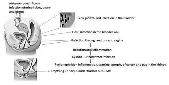
coli to cause cystitis. Do the same for pyelonephritis. Diagram the pathway taken by Neisseria gonorrhoeae to cause PID.
To draw:
Pathway taken by E. coli to cause cystitis and pyelonephritis. Pathway taken by Neisseria gonorrhoeae to cause PID
Pictorial representation:
Concept introduction:
Cystitis is a urinary tract infection, mostly caused by E coli. Bacterial growth in the urothelium of the bladder through rectum, urethra, perineum and vagina causes cystitis. Pyelonephritis is an inflammatory disease of kidneys, mostly caused by E coli. Bacteria can reach kidney through lower urinary tract and bloodstream. Pelvic inflammatory disease or PID is a severe bacterial infection of pelvic organs of women, mostly caused by N gonorrhoeae.
Explanation of Solution
Cystitis is a common urinary tract infection, caused by Escherichia coli (E coli) bacteria. Nearly 70-95% of the urinary tract infections are caused by E coli. It is a gram-negative, anaerobic, rod-shaped bacteria. It is commonly found in environment, skin and intestines of animal and human. Mostly they are harmless and help in digestion, and sometimes they can cause serious health problems like diarrhea, cystitis (urinary tract infections), respiratory problem, pneumonia, food poison and others. Cystitis is primarily caused by Escherichia coli and also by Staphylococcus saprophyticus, Klebsiella species, Proteus species and Enterobacter species. E coli covers the lining of the bladder and causes irritation and inflammation which leads to cystitis. Cystitis affects people in all age groups, however, it is more common in women than men due to the short urethra, which is less than 2 inches. The female urethra is very close to anal opening, hence it may lead to intestinal bacterial contamination. It can be prevented by drinking plenty of water which can flush bacteria and also with personal hygiene. Cystitis can be treated with antibiotics such as nitrofurantoin, trimethoprim - sulfamethoxazole, fluoroquinolone and ampicillin.
Pyelonephritis is an inflammatory disease of kidneys, caused by bacterial infection. It is a life-threatening disease which severely damages one or both kidneys and causes renal scarring. More than 70% of pyelonephritis is caused by E coli and rarely by Enterococcus faecalis, Pseudomonas aeruginosa and Klebsiella species. Bacteria can reach kidney through lower urinary tract and bloodstream. Initially, it infects bladder and move to kidney and severely damage one or both kidneys. Lower urinary tract infection is very vulnerable for kidney damage and pyelonephritis. Pyelonephritis is more common in women than men due to short urethra, which is less than 2 inches. The female urethra is very close to anal opening, hence it may lead to intestinal bacterial contaminations. Second or third generation cephalosporin is used to treat pyelonephritis.
Pelvic inflammatory disease or PID is a severe bacterial infection of pelvic organs such as uterus, cervix, uterine tubes or ovaries in female. N gonorrhoeae and C trachomatis are common microorganisms mostly responsible for PID. Uterine tubes infection or salpingitis by N gonorrhoeae leads to scarring which blocks ovary. The uterine block may cause ectopic pregnancy. Salpingitis causes10-15% infertility in women. PID can be treated with doxycycline and cefoxitin.
Cystitis is a urinary tract infection, majorly caused by E. coli leads to irritation and inflammation of the urinary bladder. Pyelonephritis is an inflammatory disease of the kidney caused by E coli. PID is an inflammatory diseases of female pelvic organ caused by N gonorrhoeae.
Want to see more full solutions like this?
Chapter 26 Solutions
Microbiology: An Introduction
Additional Science Textbook Solutions
Physical Universe
HUMAN ANATOMY
Biology: Life on Earth with Physiology (11th Edition)
Microbiology Fundamentals: A Clinical Approach


- Briefly state the physical meaning of the electrocapillary equation (Lippman equation).arrow_forwardExplain in a small summary how: What genetic information can be obtained from a Punnet square? What genetic information cannot be determined from a Punnet square? Why might a Punnet Square be beneficial to understanding genetics/inheritance?arrow_forwardIn a small summary write down:arrow_forward
- Not part of a graded assignment, from a past midtermarrow_forwardNoggin mutation: The mouse, one of the phenotypic consequences of Noggin mutationis mispatterning of the spinal cord, in the posterior region of the mouse embryo, suchthat in the hindlimb region the more ventral fates are lost, and the dorsal Pax3 domain isexpanded. (this experiment is not in the lectures).a. Hypothesis for why: What would be your hypothesis for why the ventral fatesare lost and dorsal fates expanded? Include in your answer the words notochord,BMP, SHH and either (or both of) surface ectoderm or lateral plate mesodermarrow_forwardNot part of a graded assignment, from a past midtermarrow_forward

 Medical Terminology for Health Professions, Spira...Health & NutritionISBN:9781305634350Author:Ann Ehrlich, Carol L. Schroeder, Laura Ehrlich, Katrina A. SchroederPublisher:Cengage LearningEssentials of Pharmacology for Health ProfessionsNursingISBN:9781305441620Author:WOODROWPublisher:Cengage
- Understanding Health Insurance: A Guide to Billin...Health & NutritionISBN:9781337679480Author:GREENPublisher:CengageSurgical Tech For Surgical Tech Pos CareHealth & NutritionISBN:9781337648868Author:AssociationPublisher:Cengage