Videos
Physicians’ Assistants
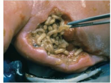
Another invertebrate animal that has been recruited for medical duty is the blowfly or, more precisely, blowfly larvae, commonly known as maggots (FIG. 24-32). Blowfly maggots have proved to be effective at ridding wounds and ulcers of dead and dying tissue. If such tissue is not removed, it can interfere with healing or lead to infection. Traditionally, dead tissue in wounds is removed by a physician wielding a scalpel, but maggots offer an increasingly common alternative treatment. In this treatment, a bandage containing day-old, sterile maggots is applied to the wound. The maggots consume dead or dying tissue, secreting digestive enzymes that do not harm healthy skin or bone. After a few days, the maggots have grown to the size of rice kernels and are removed. The treatment is repeated until the wound is clean.
FIGURE 24-32 Blowfly maggots can clean wounds
Want to see the full answer?
Check out a sample textbook solution
Chapter 24 Solutions
Biology: Life on Earth with Physiology (11th Edition)


- a) What does Wilkins's letter, (pages 2-4) add to this picture? b) What does Franklin and Gosling’s letter (pages 4-5) add to the picture? c) Why couldn’t Watson & Crick credibly publish without the inclusion of the other two letters?arrow_forwardOpen documents with docReader X 3 Listen Question 3 Determine the confidence interval (%) if there is probability value of 0.001. A 0.009% B) 0.999% ©90% LU 99% 99.9% F 9% 1 = Listen Last saved 7:05:51 PM Iarrow_forwardThe amount of drug that is administered to produce a specific effect? Dose Potencyarrow_forward
- https://www.youtube.com/watch?v=DiyWK3fzTpAarrow_forwardWhat can public health officials do about the tobacco public health problem in Indonesia? Identify one education strategy that can be taken in Indonesia.arrow_forwardIn your own words, describe the difference between weathering and erosion.arrow_forward
- In your own words, describe the difference between weathering and erosion.arrow_forwarda) Summarize the Watson & Crick model of DNA as they put forth in their letter to Nature. b) Draw a picture of Watson and Crick's proposed DNA structure, and include measurements they indicate. c) Is the model they gave still considered accurate?arrow_forwarda) Give a definition of STRs. b) Must each copy of a chromosome have the same number of repeats? c) How many STRs does the FBI use for identification in a criminal case?arrow_forward
- What part of the replication process is depicted in the photo you uploaded below? DNA polymerase I and ligase in action:arrow_forwardWhat is the purpose of each of the following steps of the DNA isolation process? Blending Salt Detergent Meat tenderizer Ice-cold isopropanolarrow_forwardUsing the envelope depiction we presented in class, draw out chemical structures for thefollowing oligonucleotides. You can abbreviate the bases as Ade, Cyt, Gua, Thy, Ura. A. d(GACA)B. p(d(TATA))C. GUCUparrow_forward

 Human Biology (MindTap Course List)BiologyISBN:9781305112100Author:Cecie Starr, Beverly McMillanPublisher:Cengage Learning
 Biology: The Unity and Diversity of Life (MindTap...BiologyISBN:9781305073951Author:Cecie Starr, Ralph Taggart, Christine Evers, Lisa StarrPublisher:Cengage Learning
 Biology 2eBiologyISBN:9781947172517Author:Matthew Douglas, Jung Choi, Mary Ann ClarkPublisher:OpenStax