
Videos
(a)
To identify: The interphase, each phase of mitosis and cytokinesis in the figure 4.4(a)-(e).
Introduction: The cell division is the process of cycle cycle in which the parent cell divided into two or more daughter cells. The cell divison is needed for growth of the individual, wound healing and replacement of old and dying cells. The duration of cell division differs on the type of the cell. The somatic cell divison consists of three phases namely interphase, mitotic phase and cytokinesis.
(a)
Answer to Problem 1.1BGL
Pictorial representation: The phase of cell cycle given in the figure 4.4 (a) is identified as interphase.
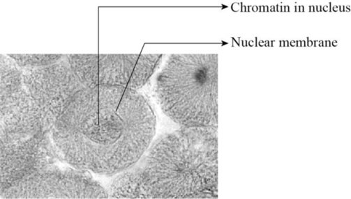
Fig 1: The interphase of somatic cell division.
Explanation of Solution
The interphase is the longest part of the cell cycle. During this phase the cell grow and replicates its DNA before moving into the mitosis. The interphase consist of three stages anmely G1 phase, S phase and G2 phase. The G1 phase is the first gap pahse in which the cell grows larger and organeeles are copied. The S phase is the synthetic phase in which the DNA of the cell replicates to produce another copy of DNA. The G2 phase is the second gap pahse in which the cells makes proteins and reorganise its contents before it enters into the mitosis.
(b)
To identify: The interphase, each phase of mitosis and cytokinesis in the figure 4.4(a)-(e).
Introduction: The cell division is the process of cycle cycle in which the parent cell divided into two or more daughter cells. The cell divison is needed for growth of the individual, wound healing and replacement of old and dying cells. The duration of cell division differs on the type of the cell. The somatic cell divison consists of three phases namely interphase, mitotic phase and cytokinesis.
(b)
Answer to Problem 1.1BGL
Pictorial representation: The phase of cell cycle given in the figure 4.4 (b) is identified as prophase.
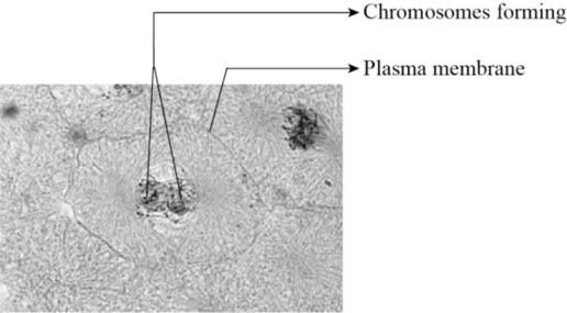
Fig 2: The prophase of somatic cell division.
Explanation of Solution
The mitotic phase of the cell division is called as M phase. This phase consists of four stages namely prophase, metaphase, anaphase and telophase. The prophase is the first phase of cell division. During this phase, the chromosomes becomes visible, the nucleolus disappears, the chromatin condenses, the mitotic spindle forms and the nuclear envelope disappears.
(c)
To identify: The interphase, each phase of mitosis and cytokinesis in the figure 4.4(a)-(e).
Introduction: The cell division is the process of cycle cycle in which the parent cell divided into two or more daughter cells. The cell divison is needed for growth of the individual, wound healing and replacement of old and dying cells. The duration of cell division differs on the type of the cell. The somatic cell divison consists of three phases namely interphase, mitotic phase and cytokinesis.
(c)
Answer to Problem 1.1BGL
Pictorial representation: The phase of cell cycle given in the figure 4.4 (c) is identified as metaphase.
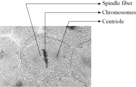
Fig 3: The metaphase of somatic cell division.
Explanation of Solution
The metaphase is the second phase of mitosis. At the start of metaphase the chromosomes are randomly arranged within the nucleus. The microtubules connects to each chromosome. Then the chromosomes are aligned in the middle of the cell with the sister chromatids of each chromosome on either side of the metaphase plate. The sister chromatids contains cohensin proteins which are broken after passing the mitotic check point to ensure alignment of chromosomes at the metaphase plate. This then acrtivates the anaphase-promoting complex which leads to end of metaphase.
(d)
To identify: The interphase, each phase of mitosis and cytokinesis in the figure 4.4(a)-(e).
Introduction: The cell division is the process of cycle cycle in which the parent cell divided into two or more daughter cells. The cell divison is needed for growth of the individual, wound healing and replacement of old and dying cells. The duration of cell division differs on the type of the cell. The somatic cell divison consists of three phases namely interphase, mitotic phase and cytokinesis.
(d)
Answer to Problem 1.1BGL
Pictorial representation: The phase of cell cycle given in the figure 4.4 (d) is identified as anaphase.
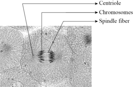
Fig 4: The anaphase of somatic cell division.
Explanation of Solution
The anaphase of cell cycle begins with the formation of anaphase promoting complex. This complex degrades the protein called securin which inhibits the separase. The separase is the protein that separates the cohesion protein that helds the chromatids together. As the result of this process the chromatids becomes apart from each other.
(e)
To identify: The interphase, each phase of mitosis and cytokinesis in the figure 4.4(a)-(e).
Introduction: The cell division is the process of cycle cycle in which the parent cell divided into two or more daughter cells. The cell divison is needed for growth of the individual, wound healing and replacement of old and dying cells. The duration of cell division differs on the type of the cell. The somatic cell divison consists of three phases namely interphase, mitotic phase and cytokinesis.
(e)
Answer to Problem 1.1BGL
Pictorial representation: The phase of cell cycle given in the figure 4.4 (e) is identified as telophase.
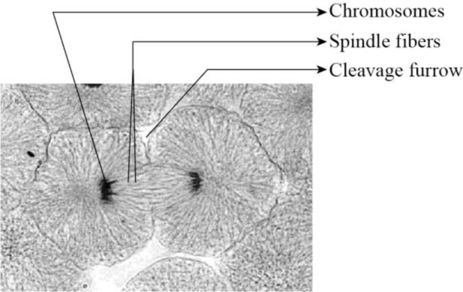
Fig 5: The telophase of somatic cell division.
Explanation of Solution
The telophase is the final stage of cell cycle. During this phase, the spindle fibers underoes depolymerisation that separstes the sister chromatids. The separate chromosomes are released from tighltly bound structure. Then the nuclear envelops reforms around the new nuclei in each half of the dividing cells.
Want to see more full solutions like this?
Chapter 4 Solutions
Laboratory Manual for Anatomy and Physiology, 6e Loose-Leaf Print Companion


- When beta-lactamase was isolated from Staphylcoccus aureus and treated with a phosphorylating agent, only the active site, serine was phosphorylated. Additionally, the serine was found to constitute 0.35% (by weight) of this beta-lactamase enzyme. Using this, calculate the molecular weight of this enzyme and estimate the number of amino acids present in the polypeptide.arrow_forwardBased on your results from the Mannitol Salt Agar (MSA) media, which of your bacteria were mannitol fermenters and which were not mannitol fermenters?arrow_forwardhelp tutor pleasearrow_forward
- Q8. A researcher wants to study the effectiveness of a pill intended to reduce stomach heartburn in pregnant women. The researcher chooses randomly 400 women to participate in this experiment for 9 months of their pregnancy period. They all need to have the same diet. The researcher designs two groups of 200 participants: One group take the real medication intended to reduce heartburn, while the other group take placebo medication. In this study what are: Independent variable: Dependent variable: Control variable: Experimental group: " Control group: If the participants do not know who is consuming the real pills and who is consuming the sugar pills. This study is It happens that 40% of the participants do not find the treatment helpful and drop out after 6 months. The researcher throws out the data from subjects that drop out. What type of bias is there in this study? If the company who makes the medication funds this research, what type of bias might exist in this research work?arrow_forwardHow do I determine the inhertiance pattern from the pedigree diagram?arrow_forwardits an open book assignemntarrow_forward
- Describe two different gene regulation mechanisms involving methylationarrow_forwardWhat is behavioral adaptarrow_forward22. Which of the following mutant proteins is expected to have a dominant negative effect when over- expressed in normal cells? a. mutant PI3-kinase that lacks the SH2 domain but retains the kinase function b. mutant Grb2 protein that cannot bind to RTK c. mutant RTK that lacks the extracellular domain d. mutant PDK that has the PH domain but lost the kinase function e. all of the abovearrow_forward
 Human Anatomy & Physiology (11th Edition)BiologyISBN:9780134580999Author:Elaine N. Marieb, Katja N. HoehnPublisher:PEARSON
 Biology 2eBiologyISBN:9781947172517Author:Matthew Douglas, Jung Choi, Mary Ann ClarkPublisher:OpenStax
 Anatomy & PhysiologyBiologyISBN:9781259398629Author:McKinley, Michael P., O'loughlin, Valerie Dean, Bidle, Theresa StouterPublisher:Mcgraw Hill Education,
 Molecular Biology of the Cell (Sixth Edition)BiologyISBN:9780815344322Author:Bruce Alberts, Alexander D. Johnson, Julian Lewis, David Morgan, Martin Raff, Keith Roberts, Peter WalterPublisher:W. W. Norton & Company
 Laboratory Manual For Human Anatomy & PhysiologyBiologyISBN:9781260159363Author:Martin, Terry R., Prentice-craver, CynthiaPublisher:McGraw-Hill Publishing Co.
 Inquiry Into Life (16th Edition)BiologyISBN:9781260231700Author:Sylvia S. Mader, Michael WindelspechtPublisher:McGraw Hill Education