Microbiology Fundamentals: A Clinical Approach
3rd Edition
ISBN: 9781260163698
Author: Cowan
Publisher: MCG
expand_more
expand_more
format_list_bulleted
Concept explainers
Textbook Question
Chapter 3, Problem 1VC
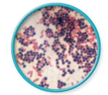
From chapter 2, figure 2.18. Explain why some cells are pink and others are purple in this image of a

Expert Solution & Answer
Want to see the full answer?
Check out a sample textbook solution
Students have asked these similar questions
C
MasteringHealth MasteringNu ×
session.healthandnutrition-mastering.pearson.com/myct/itemView?assignment ProblemID=17396416&attemptNo=1&offset=prev
10. Your instructor will give you 2 amino acids during the activity session (video 2-7.
A. First color all the polar and non-polar covalent bonds in the R groups of your 2 amino acids
using the same colors as in #7. Do not color the bonds in the backbone of each amino acid.
B. Next, color where all the hydrogen bonds, hydrophobic interactions and ionic bonds could
occur in the R group of each amino acid. Use the same colors as in #7. Do not color the bonds
in the backbone of each amino acid.
C. Position the two amino acids on the page below in an orientation where the two R groups
could bond together. Once you are satisfied, staple or tape the amino acids in place and label
the bond that you formed between the two R groups.
- Polar covalent Bond - Red
- Non polar Covalent boND- yellow
- Ionic BonD - PINK
Hydrogen Bonn - Purple
Hydrophobic interaction-green
O=C-N
H
I.
H
HO
H
=O
CH2
C-C-N
HICK
H
HO
H
CH2
OH
H₂N
C = O
Find the dental formula and enter it in the following format:
I3/3 C1/1 P4/4 M2/3 = 42 (this is not the correct number, just the correct format)
Please be aware: the upper jaw is intact (all teeth are present). The bottom jaw/mandible is not intact. The front teeth should include 6 total rectangular teeth (3 on each side) and 2 total large triangular teeth (1 on each side).
Chapter 3 Solutions
Microbiology Fundamentals: A Clinical Approach
Ch. 3.1 - List the structures all bacteria possess.Ch. 3.1 - Identify three structures some but not all...Ch. 3.1 - Describe three major shapes of bacteria.Ch. 3.1 - Provide at least four terms to describe bacterial...Ch. 3.2 - Describe the structure and function of six...Ch. 3.2 - Prob. 6AYPCh. 3.2 - Q. Device-associated infections are very common...Ch. 3.3 - Differentiate between the two main types of...Ch. 3.3 - Prob. 8AYPCh. 3.3 - Prob. 9AYP
Ch. 3.3 - Prob. 2MMCh. 3.4 - Identify seven structures that may be contained in...Ch. 3.4 - Prob. 11AYPCh. 3.4 - Prob. 1NPCh. 3.5 - Compare and contrast the major features of...Ch. 3.6 - Differentiate between Bergeys Manual of Systematic...Ch. 3.6 - Name four divisions ending in cutes and describe...Ch. 3.6 - Define a species in terms of bacteria.Ch. 3 - Archaea a. are most genetically related to...Ch. 3 - Prob. 2QCh. 3 - Suppose an argument in your city has erupted about...Ch. 3 - Prob. 4QCh. 3 - As a supervisor in the infection control unit, you...Ch. 3 - Prob. 6QCh. 3 - Prob. 7QCh. 3 - Prob. 8QCh. 3 - Bacteria and archaea have a much greater diversity...Ch. 3 - Prob. 10QCh. 3 - Bacteria have been found to change the structures...Ch. 3 - Bacterial and archaeal chromosomes are not...Ch. 3 - Prob. 13QCh. 3 - The results of your patients wound culture just...Ch. 3 - We know that bacteria/archaea and their genetics...Ch. 3 - Find the true statement about biofilms. a. They...Ch. 3 - Suggest more than one reason why bacteria may...Ch. 3 - Construct arguments agreeing with and refuting...Ch. 3 - Which of the following would be used to identify...Ch. 3 - During the cold war between the Soviet Union and...Ch. 3 - During the cold war between the Soviet Union and...Ch. 3 - From chapter 2, figure 2.18. Explain why some...
Knowledge Booster
Learn more about
Need a deep-dive on the concept behind this application? Look no further. Learn more about this topic, biology and related others by exploring similar questions and additional content below.Similar questions
- 12. Calculate the area of a circle which has a radius of 1200 μm. Give your answer in mm² in scientific notation with the correct number of significant figures.arrow_forwardDescribe the image quality of the B.megaterium at 1000X before adding oil? What does adding oil do to the quality of the image?arrow_forwardWhich of the follwowing cells from this lab do you expect to have a nucleus and why or why not? Ceratium, Bacillus megaterium and Cheek epithelial cells?arrow_forward
- 14. If you determine there to be debris on your ocular lens, explain what is the best way to clean it off without damaging the lens?arrow_forward11. Write a simple formula for converting mm to μm when the number of mm's is known. Use the variable X to represent the number of mm's in your formula.arrow_forward13. When a smear containing cells is dried, the cells shrink due to the loss of water. What technique could you use to visualize and measure living cells without heat-fixing them? Hint: you did this technique in part I.arrow_forward
- 10. Write a simple formula for converting μm to mm when the number of μm's are known. Use the variable X to represent the number of um's in your formula.arrow_forward8. How many μm² is in one cm²; express the result in scientific notation. Show your calculations. 1 cm = 10 mm; 1 mm = 1000 μmarrow_forwardFind the dental formula and enter it in the following format: I3/3 C1/1 P4/4 M2/3 = 42 (this is not the correct number, just the correct format) Please be aware: the upper jaw is intact (all teeth are present). The bottom jaw/mandible is not intact. The front teeth should include 6 total rectangular teeth (3 on each side) and 2 total large triangular teeth (1 on each side).arrow_forward
- Answer iarrow_forwardAnswerarrow_forwardcalculate the questions showing the solution including variables,unit and equations all the questiosn below using the data a) B1, b) B2, c) hybrid rate constant (1) d) hybrid rate constant (2) e) t1/2,dist f) t1/2,elim g) k10 h) k12 i) k21 j) initial concentration (C0) k) central compartment volume (V1) l) steady-state volume (Vss) m) clearance (CL) AUC (0→10 min) using trapezoidal rule n) AUC (20→30 min) using trapezoidal rule o) AUCtail (AUC360→∞) p) total AUC (using short cut method) q) volume from AUC (VAUC)arrow_forward
arrow_back_ios
SEE MORE QUESTIONS
arrow_forward_ios
Recommended textbooks for you
- Basic Clinical Lab Competencies for Respiratory C...NursingISBN:9781285244662Author:WhitePublisher:Cengage
 Microbiology for Surgical Technologists (MindTap ...BiologyISBN:9781111306663Author:Margaret Rodriguez, Paul PricePublisher:Cengage Learning
 Human Heredity: Principles and Issues (MindTap Co...BiologyISBN:9781305251052Author:Michael CummingsPublisher:Cengage Learning


Basic Clinical Lab Competencies for Respiratory C...
Nursing
ISBN:9781285244662
Author:White
Publisher:Cengage


Microbiology for Surgical Technologists (MindTap ...
Biology
ISBN:9781111306663
Author:Margaret Rodriguez, Paul Price
Publisher:Cengage Learning

Human Heredity: Principles and Issues (MindTap Co...
Biology
ISBN:9781305251052
Author:Michael Cummings
Publisher:Cengage Learning
cell culture and growth media for Microbiology; Author: Scientist Cindy;https://www.youtube.com/watch?v=EjnQ3peWRek;License: Standard YouTube License, CC-BY