
Concept explainers
Videos
To explain: The reason why the probability of death as a function of the body mass index is a U-shaped curve.
Concept introduction: The body mass index is the measure of the body fat based on the height and weight that applies to adult men and women. A body mass index between 18.5 and 25 is ideal. The body mass index between 25 and 30 is classified as overweight. The person with a BMI more than 30 is called obese. The BMI below 18 is classified as underweight person.
</SUMMARY-INTRODUCTION>
Pictorial representation: Fig.1 represents the “U” shaped curve of probability of death as a function of the body mass index.
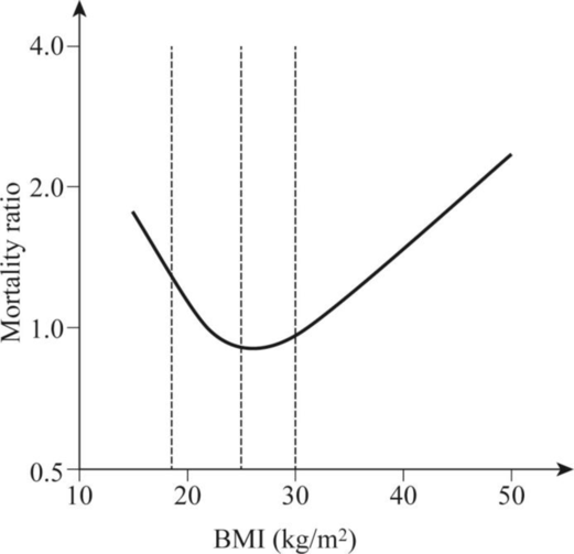
Fig. 1: “U” shaped curve of probability of death as a function of the body mass index.
Explanation:
The relationship between the all-cause mortality and body mass index in man is a U-shaped curve. The body mass index categorizes the people in overweight, normal weight, and underweight. The severely underweight individuals are at the risk of dying from the malnutrition-related causes. The individuals having 40% overweight have more chances of premature death when compared to a person with average weight. This is because obesity is linked to several serious medical conditions including diabetes and atherosclerosis. The individuals who belong to severely underweight categories and severely overweight categories are more likely to die from various reasons than the individuals who belong to normal category.
Want to see the full answer?
Check out a sample textbook solution
Chapter 22 Solutions
FUNDAMENTALS OF BIOCHEMISTRY WPNG 1-SEME


- Calculate the standard change in Gibbs free energy, AGrxn, for the given reaction at 25.0 °C. Consult the table of thermodynamic properties for standard Gibbs free energy of formation values. NH,Cl(s) →NH; (aq) + C1 (aq) AGrxn -7.67 Correct Answer Determine the concentration of NH+ (aq) if the change in Gibbs free energy, AGrxn, for the reaction is -9.27 kJ/mol. 6.49 [NH+] Incorrect Answer kJ/mol Marrow_forwardWhat are some topics of interest that neurotoxicologists study? For example, toxin-induced seizures, brain death, and such along those lines?arrow_forwardCould you help me with the explanation of the answer to exercise 15, chapter 1 of Lehinger Question Nombramiento de estereoisómeros con dos carbonos quirales utilizando el sistema RS(R,R)El isómero del metilfenidato (Ritalin) se utiliza para tratar el trastorno por déficit de atención con hiperactividad (TDAH).(S,S)El isómero es un antidepresivo. Identifique los dos carbonos quirales en la siguiente estructura. ¿Es este el(R,R)o el(S,S)¿isómero? Dibuja el otro isómero. Nombramiento de estereoisómeros con dos carbonos quirales utilizando el sistema RS(R,R)El isómero del metilfenidato (Ritalin) se utiliza para tratar el trastorno por déficit de atención con hiperactividad (TDAH).(S,S)El isómero es un antidepresivo.arrow_forward
- The reaction A+B → C + D AG°' = -7.3 kcal/mol can be coupled with which of the following unfavorable reactions to drive it forward? A. EFG+HAG° = 5.6 kcal/mol. B. J+KZ+A AG° = 2.3 kcal/mol. C. P+RY+DAG° = 8.2 kcal/mol. D. C + T → V + W AG°' = -5.9 kcal/mol. E. AN→ Q+KAG°' = 4.3 kcal/mol.arrow_forwardWhat would be the toxicological endpoints for neurotoxicity?arrow_forwardWhat are "endpoints" in toxicology exactly? Please give an intuitive easy explanationarrow_forward
- Fura-2 Fluorescence (Arbitrary Unit) 4500 4000 3500 3000 2500 2000 1500 1000 500 [Ca2+]=2970nM, 25°C [Ca2+] 2970nM, 4°C [Ca2+]=0.9nM, 25°C [Ca2+] = 0.9nM, 4°C 0 260 280 300 340 360 380 400 420 440 Wavelength (nm) ← < The figure on the LHS shows the excitation spectra of Fura-2 (Em = 510 nm) in 2 solutions with two different Ca2+ ion concentration as indicated. Except for temperature, the setting for excitation & signal acquisition was identical.< ப a) The unit in Y-axis is arbitrary (unspecified). Why? < < b) Compare & contrast the excitation wavelength of the Isosbestic Point of Fura-2 at 25 °C & 4 °C. Give a possible reason for the discrepancy. < c) The fluorescence intensity at 25 °C & 4 °C are different. Explain why with the concept of electronic configuration. <arrow_forwarddraw in the structure of each amino acid (as L-amino acids) using the Fischer projection style. an example has been included. Draw the structure for glycine, alanine, valine, isoleucine, methionine, proline, phenylalanine, tryptophan, serine, threonine, asparagine, glutamine, lysine, arginine, aspartic acid, glutamic acid, histidine, tyrosine, cysteinearrow_forwarddraw in the structure of each amino acid (as L-amino acids) using the Fischer projection style. an example has been includedarrow_forward
 BiochemistryBiochemistryISBN:9781319114671Author:Lubert Stryer, Jeremy M. Berg, John L. Tymoczko, Gregory J. Gatto Jr.Publisher:W. H. Freeman
 Lehninger Principles of BiochemistryBiochemistryISBN:9781464126116Author:David L. Nelson, Michael M. CoxPublisher:W. H. Freeman
 Fundamentals of Biochemistry: Life at the Molecul...BiochemistryISBN:9781118918401Author:Donald Voet, Judith G. Voet, Charlotte W. PrattPublisher:WILEY
 BiochemistryBiochemistryISBN:9781305961135Author:Mary K. Campbell, Shawn O. Farrell, Owen M. McDougalPublisher:Cengage Learning
 BiochemistryBiochemistryISBN:9781305577206Author:Reginald H. Garrett, Charles M. GrishamPublisher:Cengage Learning
 Fundamentals of General, Organic, and Biological ...BiochemistryISBN:9780134015187Author:John E. McMurry, David S. Ballantine, Carl A. Hoeger, Virginia E. PetersonPublisher:PEARSON