Concept explainers
Northern blot analysis is performed on cellular mRNA isolated from E. coli. The probe used in the northern blot analysis hybridizes to a portion of the lacY sequence. Below is an example of the gel from northern blot analysis for a wild
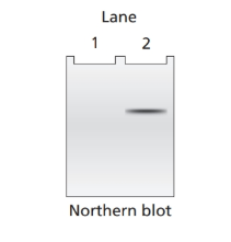
a. Lac+ bacteria with the genotype
b. Lac- bacteria with the genotype
c. Lac- bacteria with the genotype
d. Lac+ bacteria with the genotype
e. Lac- bacteria with the genotype
f. Lac- bacteria with the genotype
g. Lac- bacteria with the genotype
Want to see the full answer?
Check out a sample textbook solutionChapter 12 Solutions
Genetic Analysis: An Integrated Approach (3rd Edition)
- In this western blot, the levels of TBK remain constant with increasing amounts/expression of the viral protein (VP). Figure description: Increasing amounts of a plasmid expressing the viral protein (0.5, 1, or 2ug) were cotransfected with TBK1 expression plasmid. Cells were harvested 24 h post-transfection and analyzed for phosphorylated TBK1 (anti-pTBK1 Ser172), total TBK1 (anti-TBK1), B-actin (anti-B-actin), and viral protein (anti-VP) expression by Western blot analysis. VP PTBK1 (S172) TBK1 actin Virus proteins O True O Falsearrow_forwardDNA samples from four individuals were cleaved with the same MW restriction endonuclease. The DNA fragments were separated by gel clectrophoresis, transferred to a membrane, and hybridized with a 12 kb 10 kb DNA probe complementary to a region between sites C and D (see 8 kb hybridization line). The image of the southern blot shows the labeled DNA bands and 6 kb molecular weight (MW) markers. The lane labels I, II, III, and IV -5 kb correspond to individuals I, II, III, and IV. Assume that fragments such as C-D and C-E are clearly resolved in this gel system. Fragment sizes are as given: A-B is 4 kb, B-C is I kb, C-D is 5 kb, and D-E is 650 bp. Individual I has five cleavage sites (A, B, C, D, and E) for the restriction endonuclease. DNA homologous to probe Which individual has at least one point mutation that eliminates restriction site C only? O II IV cannot be determined II Which individual has at least one point mutation that climinates restriction sites B and C? III O IV cannot be…arrow_forwardAfter characterizing the DNA composition of various cats, you identify a protein-coding gene in tigers called stripes and wish to study the structure of the protein product STRIPES. This requires that you purify recombinant ridges from E. coli. First, the stripes gene must be amplified by PCR and then inserted into an appropriate plasmid for bacterial expression. Such a plasmid is diagrammed below. ori CAP Binding Site من Promoter MCS The restriction sites for Aatll and Kpnl are: Aatll 5'-GACGTC-3' Kpnl = 5'-GGTACC-3' Laco (Operator) -Kpnl Aatll The coding strand for the stripes gene is shown below, with start and stop codons in bold. 5'-ATGCAACAGTAGCTGAAGCCCAGTGACACCATCGAAAATGTGAAGGCCAAGATGAGGCTCATCTTTGCAGGCAAGCAGCTG GAAGATGGCCGTACTCTTTCTGACTATGCGTCTGAGAGGTGGTATGCAGATCTTCGTGAAGACCCTGACCGGCAAGACCAATGT GAAGGCCAAGATCCAGGATAAAGAAGGCATCCCTCCCGACCAGCAGAGGGCACTCTTTCTGACTACAACATCCAGAAGGAGTCG ACCCTGCACCTGGTCCTGCTGACCGGCAAGACCATCACTCTGGAGGTGGAGCCCAGTGACACCATCGAAAATCCCGACCAGCAG…arrow_forward
- Analysis of a 49kDA protein is aimed with a western blot technique. For this purpose, "whole cell extract" was obtained from biopsy samples taken from three different individuals, and when their spectrophotometric measurements were made, their concentrations were determined to be 40 µg/µL, 50 µg/µL, and 100 µg/µL for three samples, respectively. At the beginning of the experiment, 200 µg of protein is loaded into each gel well by using a ready 5x loading solution containing SDS and β-mercapta-ethanol together with glycerol and methylene blue dye. a) If loading will be done in a volume of 20µL, then how is the preparation of the samples before loading?b) Considering the loading content given above, what can be said about whether this PAGE is "non-denaturing" or "denaturing". Justify taking into account the loading solution content.arrow_forwardYou are attempting to prepare a single gene knockout library using the pRL27 transposon system. You grow the donor E. coli in Luria broth containing both kanamycin and diaminopimelic acid (DAP) and your recipient Serratia rubidaea in plain Luria broth. You combine an equal ratio of donor and recipient cultures and plate the mixture onto Luria agar supplemented with DAP. After 24 hours incubation at 37°C, you create a cell slurry and plate the cells onto Luria agar aupplemented with kanamycin. After 24 hours incubation at 37°C, you find that no colonies grow. What best explains this outcome? A. Failure to supplement media with DAP B. Failure to remove antiobiotic containing media C. Failure to incubate for a sufficient length of time D. Failure to incubate at the appropiate temperature E. Failure to use the proper mating mix ratioarrow_forward7. Consider the following plasmid (size 6700 bp), with restriction sites at the positions indicated: BamHI + 1 6700 bp 2800 3500 EcoRI BamHI Probearrow_forward
- Decide on two restriction sites that you can use to clone this into pL4440’s MCS. Identify their sequence and explain why. Tip: The plasmid map is showed below, details of restriction site sequences can be found at https://enzymefinder.neb.com/#!#nebheaderarrow_forwardMany resistance mechanisms are encoded on plasmids. These mechanisms are of great clinical significance, because they can spread very easily through horizontal gene transfer. A culture of the bacterial isolate is grown, and plasmid DNA is isolated using a spin column-based solid phase extraction method. The purified plasmid DNA is then submitted for next-generation sequencing. Bioinformatic analyses of the sequencing results suggests that the following gene is likely involved in antibiotic resistance: > putative antibiotic resistance gene ATGCGTGTATTAGCCTTATCGGCTGTGTTTTTGGTGGCATCGATT ATCGGAATGCCTGCGGTAGCAAAGGAATGGCAAGAAAACAAAAGT TGGAATGCTCACTTTACTGAACATAAATCACAGGGCGTAGTTGTG CTCTGGAATGAGAATAAGCAGCAAGGATTTACCAATAATCTTAAA CGGGCGAACCAAGCATTTTTACCCGCATCTAGTGCGAAAATTCCC AATAGCTTGATCGCCCTCGATTTGGGCGTGGTTAAGGATGAACAC CAAGTCTTTAAGTGGGATGGACAGACGCGCGATATCGCCACTTGG AATCGCGATCATAATCTAATCACCGCGATGAAATATTCAGTTGTG CCTGTTTATCAAGAATTTGCCCGCCAAATTGGCGAGGCACGTATG…arrow_forwardThe map of plasmid pUC19 is shown below. The restriction site coordinate is the position of the 5’base on the top strand of each site sequence. The restriction enzyme sites are in bold type if there is only one site in pUC19. Please list the fragments in order of size, largest to smallest, which will result from a complete digestion by the restriction enzyme PvuII. Please list the fragments in order of size, largest to smallest, which will result from a complete digestion by the restriction enzyme DrdI.arrow_forward
- Genomic DNA from a family where sickle-cell disease is known to be hereditary, is digested with the restriction enzyme MstII and run in a Southern Blot. The blot is hybridised with two different 0.6 kb probes, both probes (indicated in red in the diagram below) are specific for the β-globin gene (indicated as grey arrow on the diagram below). The normal wild-type βA allele contains an MstII restriction site indicated with the asterisk (*) in the diagram below; in the mutated sickle-cell βS allele this restriction site has been lost. What size bands would you expect to see on the Southern blots using probe 1 and probe 2 for an individual with sickle cell disease (have 2 βS alleles)? Probe 1 Probe 2 (a) 0.6kb 0.6kb and 1.2kb (b) 0.6kb and 1.8kb 0.6kb, 1.2kb and 1.8kb (c) 1.2kb 0.6kb (d) 1.8kb 1.8kb a. (a) b. (b) c. (c) d. (d)arrow_forwardMouse genomic DNA is treated with a restriction endonuclease and electrophoresed in an agarose gel. A radioactive probe made from the human gene rxr-1 is used to perform a Southern blot. The experiment was repeated three times. Explain the results of these repeated experiments:arrow_forwardThe right-hand column contains descriptions of 5 types of molecular markers. Label each description with the name of the being described. Single-nucleotide polymorphism (SNP) Sequence-tagged site (STS) Microsatellite Amplified restriction fragment length polymorphism (AFLP) Restriction fragment length polymorphism (RFLP) Reset Zoom TABLE 23.1 Common Types of Molecular Markers Marker Description A site in a genome where the distance between two restriction sites varies among different individuals. These sites are identified by restriction enzyme digestion of chromosomal DNA and the use of Southern blotting. Amplified restriction fragment length polymorphism (AFLP) The same as an RFLP except that the site is amplified via PCR instead of isolating the chromosomal DNA. A site in the genome that contains many short sequences that are repeated many times in a row. The total length is usually in the range of 50-200 bp, and the length of a given microsatellite may be polymorphic within a…arrow_forward
- Human Anatomy & Physiology (11th Edition)BiologyISBN:9780134580999Author:Elaine N. Marieb, Katja N. HoehnPublisher:PEARSONBiology 2eBiologyISBN:9781947172517Author:Matthew Douglas, Jung Choi, Mary Ann ClarkPublisher:OpenStaxAnatomy & PhysiologyBiologyISBN:9781259398629Author:McKinley, Michael P., O'loughlin, Valerie Dean, Bidle, Theresa StouterPublisher:Mcgraw Hill Education,
- Molecular Biology of the Cell (Sixth Edition)BiologyISBN:9780815344322Author:Bruce Alberts, Alexander D. Johnson, Julian Lewis, David Morgan, Martin Raff, Keith Roberts, Peter WalterPublisher:W. W. Norton & CompanyLaboratory Manual For Human Anatomy & PhysiologyBiologyISBN:9781260159363Author:Martin, Terry R., Prentice-craver, CynthiaPublisher:McGraw-Hill Publishing Co.Inquiry Into Life (16th Edition)BiologyISBN:9781260231700Author:Sylvia S. Mader, Michael WindelspechtPublisher:McGraw Hill Education