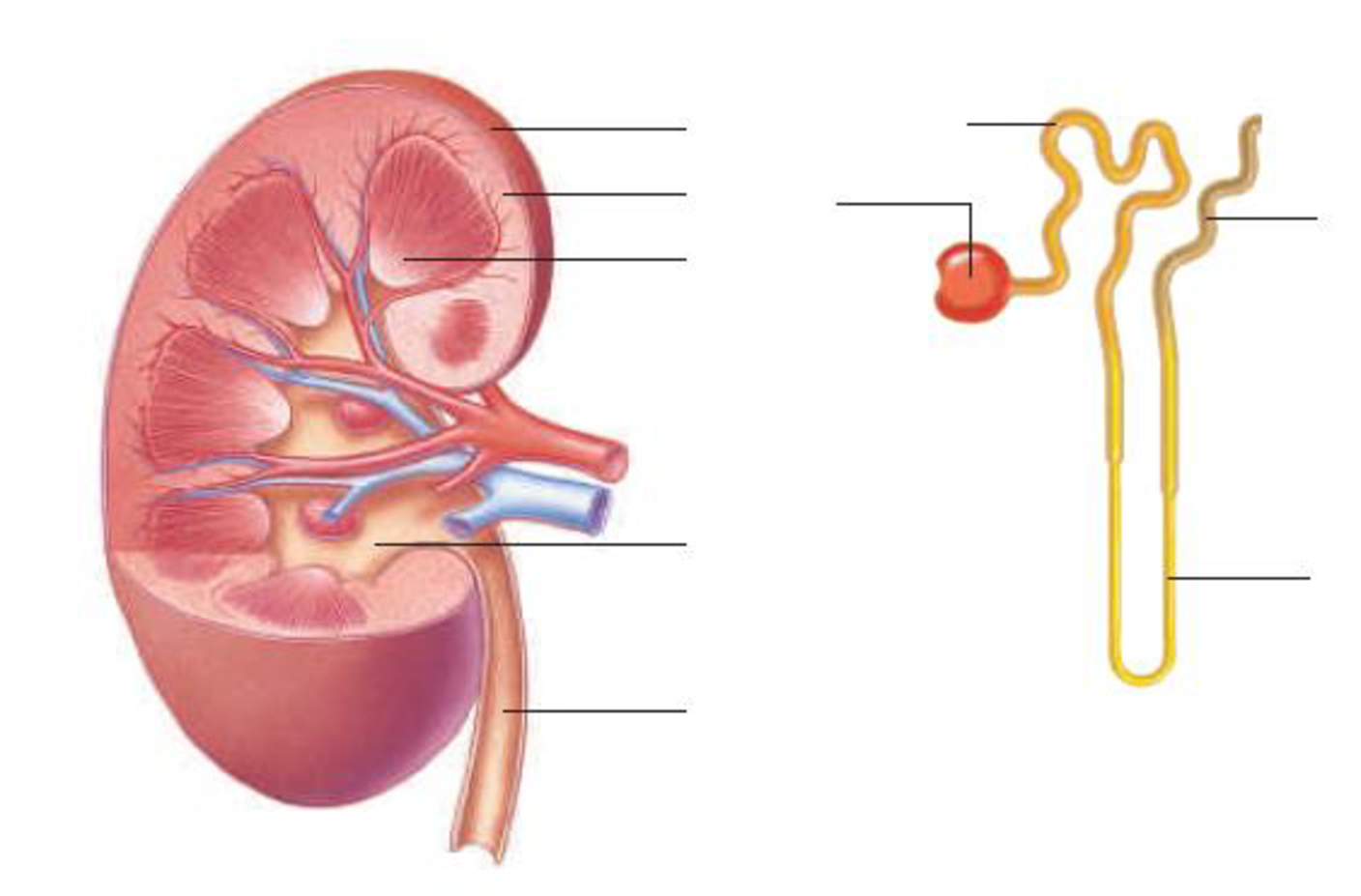
Label the parts of this kidney and nephron.

To label: The parts of the kidney and nephron.
Introduction: The kidney is an important organ of the excretory system, and nephron is the basic structural unit of the kidney. As blood passes through the kidneys, it filters the nitrogenous waste to form urine. It maintains homeostasis of the body.
Answer to Problem 1RQ
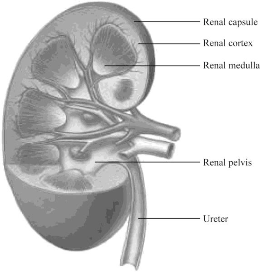
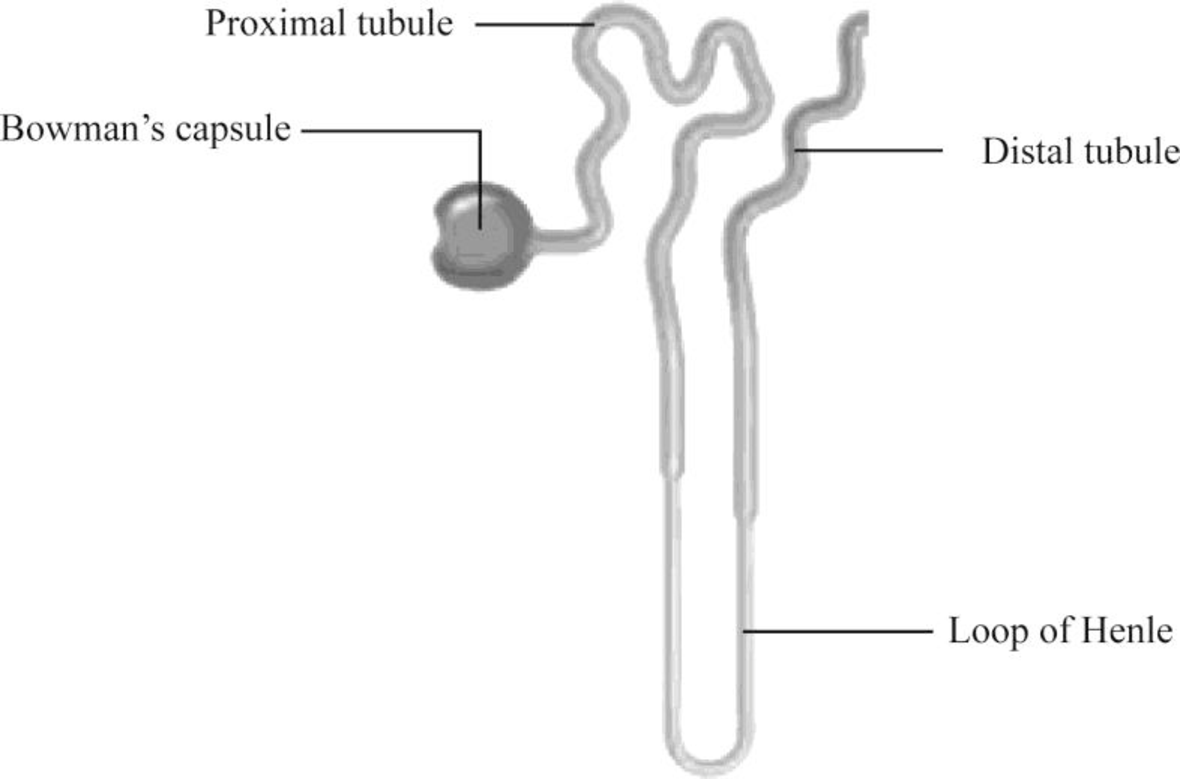
Pictorial representation: Fig.1 shows the parts of the kidney, and Fig.2 shows the parts of the nephron.

Fig.1: Kidney

Fig.2: Nephron
Explanation of Solution
Structure of a kidney:
The kidney has two zones namely renal cortex and renal medulla. Renal cortex is the outer part of kidney. It consists of blood vessels that are connected to nephrons. Renal medulla is the inner part of the kidney containing 8–12 renal pyramids consisting of collecting tubules. Each kidney has a medially located space called renal sinus. This space is the urine drainage area. It is organized into minor calyces, major calyces, and a renal pelvis. There are 8 to 15 funnel-shaped minor calyces that are associated with the renal pyramid. Several minor calyces merge to form large major calyces. There are two to three major calyces in each kidney. The major calyces merge to form a large renal pelvis. Renal pelvis merges with the ureter at the medial edge of the kidneys.
Structure of a nephron:
Nephron is the functional filtration unit of kidney. It is microscopic in nature. Nephron has divided into renal corpuscle and renal tubule. Renal tubule is composed of simple epithelium that rests on the basement membrane. Renal tubule extends from the glomerular capsule (also called Bowman's capsule. It collects the material obtained from glomeruli) and ends at the collecting duct. It consists of three continuous sections. They are as follows:
- • The proximal convoluted tubule
- • The nephron loop
- • The distal convoluted tubule
The convoluted tubules reside in the cortex, but the nephron loop usually extends into the medulla from the cortex.
The proximal convoluted tubule (PCT) is the first region of the renal tubule that is originated from the tubular pole of renal capsule. It consists of simple cuboidal epithelium and is marked by tall, apical microvilli that increase its surface area and reabsorption capacity. Under light microscope, the lumen of the proximal convoluted tubule looks fuzzy due to microvilli.
The nephron loop originates at the sharp bend in the proximal convoluted tubules. Each nephron has two limbs, namely descending limb and ascending limb. Both limbs are continuous at a hairpin within the medulla. From the proximal convoluted tubule, the descending limb extends medially to the tip of the nephron loop. Ascending limb of the nephron loop returns to the renal cortex and terminates at the distal convoluted tubule. Some parts of these limbs are classified as thick or thin according to the epithelia composing them. Thick segments of each limb have a simple cuboidal epithelium lining, whereas the thin segments of each limb have a simple squamous epithelium.
The distal convoluted tubule (DCT) originates in the renal cortex at the end of the nephron loop’s thick ascending limb and extends to a collecting tubule. DCT is composed of simple cuboidal epithelium. DCT is smaller and has only sparse, short, apical microvilli. DCT does not look fuzzy when viewed under the light microscope.
Want to see more full solutions like this?
Chapter 12 Solutions
EBK HUMAN BIOLOGY
- You intend to insert patched dominant negative DNA into the left half of the neural tube of a chick. 1) Which side of the neural tube would you put the positive electrode to ensure that the DNA ends up on the left side? 2) What would be the internal (within the embryo) control for this experiment? 3) How can you be sure that the electroporation method itself is not impacting the embryo? 4) What would you do to ensure that the electroporation is working? How can you tell?arrow_forwardDescribe a method to document the diffusion path and gradient of Sonic Hedgehog through the chicken embryo. If modifying the protein, what is one thing you have to consider in regards to maintaining the protein’s function?arrow_forwardThe following table is from Kumar et. al. Highly Selective Dopamine D3 Receptor (DR) Antagonists and Partial Agonists Based on Eticlopride and the D3R Crystal Structure: New Leads for Opioid Dependence Treatment. J. Med Chem 2016.arrow_forward
- The following figure is from Caterina et al. The capsaicin receptor: a heat activated ion channel in the pain pathway. Nature, 1997. Black boxes indicate capsaicin, white circles indicate resinferatoxin. You are a chef in a fancy new science-themed restaurant. You have a recipe that calls for 1 teaspoon of resinferatoxin, but you feel uncomfortable serving foods with "toxins" in them. How much capsaicin could you substitute instead?arrow_forwardWhat protein is necessary for packaging acetylcholine into synaptic vesicles?arrow_forward1. Match each vocabulary term to its best descriptor A. affinity B. efficacy C. inert D. mimic E. how drugs move through body F. how drugs bind Kd Bmax Agonist Antagonist Pharmacokinetics Pharmacodynamicsarrow_forward
- 50 mg dose of a drug is given orally to a patient. The bioavailability of the drug is 0.2. What is the volume of distribution of the drug if the plasma concentration is 1 mg/L? Be sure to provide units.arrow_forwardDetermine Kd and Bmax from the following Scatchard plot. Make sure to include units.arrow_forwardChoose a catecholamine neurotransmitter and describe/draw the components of the synapse important for its signaling including synthesis, packaging into vesicles, receptors, transporters/degradative enzymes. Describe 2 drugs that can act on this system.arrow_forward
- The following figure is from Caterina et al. The capsaicin receptor: a heat activated ion channel in the pain pathway. Nature, 1997. Black boxes indicate capsaicin, white circles indicate resinferatoxin. a) Which has a higher potency? b) Which is has a higher efficacy? c) What is the approximate Kd of capsaicin in uM? (you can round to the nearest power of 10)arrow_forwardWhat is the rate-limiting-step for serotonin synthesis?arrow_forwardWhat enzyme is necessary for synthesis of all of the monoamines?arrow_forward
 Human Biology (MindTap Course List)BiologyISBN:9781305112100Author:Cecie Starr, Beverly McMillanPublisher:Cengage Learning
 Biology 2eBiologyISBN:9781947172517Author:Matthew Douglas, Jung Choi, Mary Ann ClarkPublisher:OpenStax


