
Laboratory Manual For Human Anatomy & Physiology
4th Edition
ISBN: 9781260159363
Author: Martin, Terry R., Prentice-craver, Cynthia
Publisher: McGraw-Hill Publishing Co.
expand_more
expand_more
format_list_bulleted
Videos
Textbook Question
Chapter 35, Problem F35.14A
Partial frontal cut of dissected cow eye. Label the internal structures using the list provided.
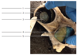
Ciliary body Sclera
Lens Tapetumfibrosum (of choroid)
Retina
McGraw. Hill Education/Cynthia Prentice-Craver
Expert Solution & Answer
Want to see the full answer?
Check out a sample textbook solution
Students have asked these similar questions
Identify the indicated cavity (Fucus).
a. antheridia
b. conceptacel
c. receptacle
d. oogonium
e. none of these
Identify the indicated structure (Saprolegnia).
a. antheridium
O b. oospore
c.sperm
d. auxospore
e. tetraspore
Of. zygospore
Using information from the primary literature (several references have been provided as a starting point below) please answer the following question: Based on your review of the literature on rewilding, what are the major scientific pros and cons for rewilding?
Please note that the focus of this assignment are the (biological) scientific issues associated with rewilding. As will be discussed in class, there are a number of non-scientific issues involved or implicated in rewilding, all ultimately affecting the public acceptability of rewilding. Although these issues are important – indeed, critical – in this assignment you should focus on the biological science issues and questions.
Details:
You must enumerate at least two pros and at least two cons.
Your answer should be no more than 500 well-chosen words, excluding references. Think carefully about how best to organize and structure your answer. Aim for high information density: say a lot, but say it succinctly. Recall Nietzche’s…
Chapter 35 Solutions
Laboratory Manual For Human Anatomy & Physiology
Ch. 35 - The cornea and the sclera compose the ______ layer...Ch. 35 - We are able to see color because the eye contains...Ch. 35 - The perception of vision occurs in the a. optic...Ch. 35 - Which of the following is not part of the middle...Ch. 35 - The area of our eye where visual acuity is best is...Ch. 35 - Which of the following extrinsic skeletal muscles...Ch. 35 - The conjunctiva covers the superficial surface of...Ch. 35 - Tears from the lacrimal gland eventually flow...Ch. 35 - Figure 35.12 Label the structures in the sagittal...Ch. 35 - FIGURE 35.13 Sagittal of the eyes (5*). Identify...
Ch. 35 - Match the terms in column A with the descriptions...Ch. 35 - Prob. 2.13ACh. 35 - List three ways in which rods and cones differ in...Ch. 35 - Partial frontal cut of dissected cow eye. Label...Ch. 35 - Prob. 3.1ACh. 35 - What kind of tissue do you think is responsible...Ch. 35 - How do you compare the shape of the pupil in the...Ch. 35 - Where was the aqueous humor in the dissected eye?Ch. 35 - What is the function of the dark pigment in the...Ch. 35 - Prob. 3.6ACh. 35 - Describe the vitreous humor of the dissected eye.Ch. 35 - A song blow to the head might cause the retina to...
Knowledge Booster


Learn more about
Need a deep-dive on the concept behind this application? Look no further. Learn more about this topic, biology and related others by exploring similar questions and additional content below.Similar questions
- Using information from the primary literature (several references have been provided as a starting point below) please answer the following question: Based on your review of the literature on rewilding, what are the major scientific pros and cons for rewilding? Please note that the focus of this assignment are the (biological) scientific issues associated with rewilding. As will be discussed in class, there are a number of non-scientific issues involved or implicated in rewilding, all ultimately affecting the public acceptability of rewilding. Although these issues are important – indeed, critical – in this assignment you should focus on the biological science issues and questions. Details: You must enumerate at least two pros and at least two cons. Your answer should be no more than 500 well-chosen words, excluding references. Think carefully about how best to organize and structure your answer. Aim for high information density: say a lot, but say it succinctly. Recall Nietzche’s…arrow_forwardNow draw a rough sketch of what the control data might look like if in addition to the specific binding, there was also a considerable amount of nonspecific binding (again using a normal dose/response curve) (do % total bound ligand vs concentration)arrow_forwardWhat are functions of cuboidal cells in the kidney? Select all that apply. Concentration of gases Dilution of chemicals Secretion of molecules Nutrition to tissues Support of tissues Absorption of moleculesarrow_forward
- question1 In plants, epithelial tissue is only found as the outermost cell layer and acts as a barrier. In humans, epithelial tissue is found inside the body as well as on the surface. What function(s) does/do epithelial tissue carry out in humans? Select all that apply. Waste storage Filtration Oxygen transport Protection Diffusion Osmosis Absorptionarrow_forwardWhat words best describes this organism? a. Unicellular/nonmotile Ob. unicellular/motile c. colonial/nonmotile d. colonial/motile e. multicelluar O f. siphonous g. none of thesearrow_forwardIdentify the phylum or class. a. Euglenophyta b. Dinoflagellata c. Bacillariophyceae d. Oomycetes e. Phaeophyceae O f. Myxomycota g. Xanthophyceae ○ h. Chrysophyceae i. Dictyosteliomycota O j. Rhodophyta Ok. Chlorophyceaens I. Charophyceaensarrow_forward
- What is produced inside the indicated structure (Fucus). a. eggs O b. antheridia ○ c. sperm d. zygotes e. none of thesearrow_forwardGreen Algae, as a group, is actually paraphyletic with one subgroup more closely related to higher plants than the other. Which of the following green algae groups is more closely related to higher plants: a. Charophyceans b. Chlorophyceans c. Rhodophyta d. Xanthophyceansarrow_forwardA single-celled green algal genus that is motile with 2 flagella, has a cup shaped chloroplast, and an eyespot: a. Volvox b. Chlamydomonas c. Euglena d. Codiumarrow_forward
- A[n] ___ is produced by members of the Myxomycota when there is a lack of moisture. a. plasmodiocarp b. aethalium c. sclerotium d. plasmodiumarrow_forwardWhich of the following is not true about the life-cycle of Fucus. a. 8 eggs per oogonium b. 64 sperm per antheridium c. eggs are flagellated d. sperm are flagellatedarrow_forwardGreen Algae, as a group, is actually paraphyletic with one subgroup more closely related to higher plants than the other. Which of the following green algae groups is more closely related to higher plants: a. Charophyceans b. Chlorophyceans c. Rhodophyta d. Xanthophyceansarrow_forward
arrow_back_ios
SEE MORE QUESTIONS
arrow_forward_ios
Recommended textbooks for you
 Medical Terminology for Health Professions, Spira...Health & NutritionISBN:9781305634350Author:Ann Ehrlich, Carol L. Schroeder, Laura Ehrlich, Katrina A. SchroederPublisher:Cengage Learning
 Understanding Health Insurance: A Guide to Billin...Health & NutritionISBN:9781337679480Author:GREENPublisher:Cengage
- Essentials of Pharmacology for Health ProfessionsNursingISBN:9781305441620Author:WOODROWPublisher:Cengage
Medical Terminology for Health Professions, Spira...
Health & Nutrition
ISBN:9781305634350
Author:Ann Ehrlich, Carol L. Schroeder, Laura Ehrlich, Katrina A. Schroeder
Publisher:Cengage Learning
Understanding Health Insurance: A Guide to Billin...
Health & Nutrition
ISBN:9781337679480
Author:GREEN
Publisher:Cengage
Essentials of Pharmacology for Health Professions
Nursing
ISBN:9781305441620
Author:WOODROW
Publisher:Cengage
Complications during Labour and Delivery; Author: FirstCry Parenting;https://www.youtube.com/watch?v=QnCviG4GpYg;License: Standard YouTube License, CC-BY