
Laboratory Manual For Human Anatomy & Physiology
4th Edition
ISBN: 9781260159363
Author: Martin, Terry R., Prentice-craver, Cynthia
Publisher: McGraw-Hill Publishing Co.
expand_more
expand_more
format_list_bulleted
Concept explainers
Videos
Textbook Question
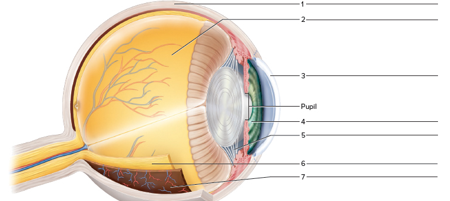
Chapter 35, Problem F35.12A
Figure 35.12 Label the structures in the sagittal section of the eye.
Expert Solution & Answer
Want to see the full answer?
Check out a sample textbook solution
Students have asked these similar questions
what key characteristics would you look for when identifying microbes?
If you had an unknown microbe, what steps would you take to determine what type of microbe (e.g., fungi, bacteria, virus) it is? Are there particular characteristics you would search for? Explain.
avorite Contact
avorite Contact
favorite Contact
୫
Recant Contacts
Keypad
Messages
Pairing
ง
107.5
NE
Controls
Media Apps Radio
Nav Phone
SCREEN
OFF
Safari File Edit View History Bookmarks Window Help
newconnect.mheducation.com
M Sign in...
S The Im...
QFri May 9 9:23 PM
w The Im...
My first....
Topic:
Mi Kimberl
M Yeast F
Connection lost! You are not connected to internet
Sigh in...
Sign in...
The Im...
S Workin...
The Im.
INTRODUCTION
LABORATORY SIMULATION
Tube 1
Fructose)
esc
- X
Tube 2
(Glucose)
Tube 3
(Sucrose)
Tube 4
(Starch)
Tube 5
(Water)
CO₂ Bubble Height (mm)
How to Measure
92
3
5
6
METHODS
RESET
#3
W
E
80
A
S
D
9
02
1
2
3
5
2
MY NOTES
LAB DATA
SHOW LABELS
%
5
T
M dtv
96
J:
ப
27
כ
00
alt
A
DII
FB
G
H
J
K
PHASE 4:
Measure gas bubble
Complete the following steps:
Select ruler and place next to tube
1. Measure starting height of gas
bubble in respirometer 1. Record in
Lab Data
Repeat measurement for tubes 2-5
by selecting ruler and move next to
each tube. Record each in Lab
Data…
Chapter 35 Solutions
Laboratory Manual For Human Anatomy & Physiology
Ch. 35 - The cornea and the sclera compose the ______ layer...Ch. 35 - We are able to see color because the eye contains...Ch. 35 - The perception of vision occurs in the a. optic...Ch. 35 - Which of the following is not part of the middle...Ch. 35 - The area of our eye where visual acuity is best is...Ch. 35 - Which of the following extrinsic skeletal muscles...Ch. 35 - The conjunctiva covers the superficial surface of...Ch. 35 - Tears from the lacrimal gland eventually flow...Ch. 35 - Figure 35.12 Label the structures in the sagittal...Ch. 35 - FIGURE 35.13 Sagittal of the eyes (5*). Identify...
Ch. 35 - Match the terms in column A with the descriptions...Ch. 35 - Prob. 2.13ACh. 35 - List three ways in which rods and cones differ in...Ch. 35 - Partial frontal cut of dissected cow eye. Label...Ch. 35 - Prob. 3.1ACh. 35 - What kind of tissue do you think is responsible...Ch. 35 - How do you compare the shape of the pupil in the...Ch. 35 - Where was the aqueous humor in the dissected eye?Ch. 35 - What is the function of the dark pigment in the...Ch. 35 - Prob. 3.6ACh. 35 - Describe the vitreous humor of the dissected eye.Ch. 35 - A song blow to the head might cause the retina to...
Knowledge Booster


Learn more about
Need a deep-dive on the concept behind this application? Look no further. Learn more about this topic, biology and related others by exploring similar questions and additional content below.Similar questions
- Ch.23 How is Salmonella able to cross from the intestines into the blood? A. it is so small that it can squeeze between intestinal cells B. it secretes a toxin that induces its uptake into intestinal epithelial cells C. it secretes enzymes that create perforations in the intestine D. it can get into the blood only if the bacteria are deposited directly there, that is, through a puncture — Which virus is associated with liver cancer? A. hepatitis A B. hepatitis B C. hepatitis C D. both hepatitis B and C — explain your answer thoroughlyarrow_forwardCh.21 What causes patients infected with the yellow fever virus to turn yellow (jaundice)? A. low blood pressure and anemia B. excess leukocytes C. alteration of skin pigments D. liver damage in final stage of disease — What is the advantage for malarial parasites to grow and replicate in red blood cells? A. able to spread quickly B. able to avoid immune detection C. low oxygen environment for growth D. cooler area of the body for growth — Which microbe does not live part of its lifecycle outside humans? A. Toxoplasma gondii B. Cytomegalovirus C. Francisella tularensis D. Plasmodium falciparum — explain your answer thoroughlyarrow_forwardCh.22 Streptococcus pneumoniae has a capsule to protect it from killing by alveolar macrophages, which kill bacteria by… A. cytokines B. antibodies C. complement D. phagocytosis — What fact about the influenza virus allows the dramatic antigenic shift that generates novel strains? A. very large size B. enveloped C. segmented genome D. over 100 genes — explain your answer thoroughlyarrow_forward
- What is this?arrow_forwardMolecular Biology A-C components of the question are corresponding to attached image labeled 1. D component of the question is corresponding to attached image labeled 2. For a eukaryotic mRNA, the sequences is as follows where AUGrepresents the start codon, the yellow is the Kozak sequence and (XXX) just represents any codonfor an amino acid (no stop codons here). G-cap and polyA tail are not shown A. How long is the peptide produced?B. What is the function (a sentence) of the UAA highlighted in blue?C. If the sequence highlighted in blue were changed from UAA to UAG, how would that affecttranslation? D. (1) The sequence highlighted in yellow above is moved to a new position indicated below. Howwould that affect translation? (2) How long would be the protein produced from this new mRNA? Thank youarrow_forwardMolecular Biology Question Explain why the cell doesn’t need 61 tRNAs (one for each codon). Please help. Thank youarrow_forward
- Molecular Biology You discover a disease causing mutation (indicated by the arrow) that alters splicing of its mRNA. This mutation (a base substitution in the splicing sequence) eliminates a 3’ splice site resulting in the inclusion of the second intron (I2) in the final mRNA. We are going to pretend that this intron is short having only 15 nucleotides (most introns are much longer so this is just to make things simple) with the following sequence shown below in bold. The ( ) indicate the reading frames in the exons; the included intron 2 sequences are in bold. A. Would you expected this change to be harmful? ExplainB. If you were to do gene therapy to fix this problem, briefly explain what type of gene therapy youwould use to correct this. Please help. Thank youarrow_forwardMolecular Biology Question Please help. Thank you Explain what is meant by the term “defective virus.” Explain how a defective virus is able to replicate.arrow_forwardMolecular Biology Explain why changing the codon GGG to GGA should not be harmful. Please help . Thank youarrow_forward
- Stage Percent Time in Hours Interphase .60 14.4 Prophase .20 4.8 Metaphase .10 2.4 Anaphase .06 1.44 Telophase .03 .72 Cytukinesis .01 .24 Can you summarize the results in the chart and explain which phases are faster and why the slower ones are slow?arrow_forwardCan you circle a cell in the different stages of mitosis? 1.prophase 2.metaphase 3.anaphase 4.telophase 5.cytokinesisarrow_forwardWhich microbe does not live part of its lifecycle outside humans? A. Toxoplasma gondii B. Cytomegalovirus C. Francisella tularensis D. Plasmodium falciparum explain your answer thoroughly.arrow_forward
arrow_back_ios
SEE MORE QUESTIONS
arrow_forward_ios
Recommended textbooks for you
 Human Physiology: From Cells to Systems (MindTap ...BiologyISBN:9781285866932Author:Lauralee SherwoodPublisher:Cengage Learning
 Human Biology (MindTap Course List)BiologyISBN:9781305112100Author:Cecie Starr, Beverly McMillanPublisher:Cengage Learning

 Principles Of Radiographic Imaging: An Art And A ...Health & NutritionISBN:9781337711067Author:Richard R. Carlton, Arlene M. Adler, Vesna BalacPublisher:Cengage Learning
 Biology (MindTap Course List)BiologyISBN:9781337392938Author:Eldra Solomon, Charles Martin, Diana W. Martin, Linda R. BergPublisher:Cengage Learning
Human Physiology: From Cells to Systems (MindTap ...
Biology
ISBN:9781285866932
Author:Lauralee Sherwood
Publisher:Cengage Learning
Human Biology (MindTap Course List)
Biology
ISBN:9781305112100
Author:Cecie Starr, Beverly McMillan
Publisher:Cengage Learning
Principles Of Radiographic Imaging: An Art And A ...
Health & Nutrition
ISBN:9781337711067
Author:Richard R. Carlton, Arlene M. Adler, Vesna Balac
Publisher:Cengage Learning
Biology (MindTap Course List)
Biology
ISBN:9781337392938
Author:Eldra Solomon, Charles Martin, Diana W. Martin, Linda R. Berg
Publisher:Cengage Learning
Anatomical Position And Directional Terms - Anatomical Terms - Directional Terms Anatomy; Author: Whats Up Dude;https://www.youtube.com/watch?v=pQUMJ6Gh9Bw;License: Standard YouTube License, CC-BY