Biology: The Unity and Diversity of Life (MindTap Course List)
14th Edition
ISBN: 9781305073951
Author: Cecie Starr, Ralph Taggart, Christine Evers, Lisa Starr
Publisher: Cengage Learning
expand_more
expand_more
format_list_bulleted
Concept explainers
Videos
Textbook Question
Chapter 31, Problem 4CT
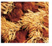
The micrograph to the right shows cells from the lining of an airway leading to the lungs. The gold cells are ciliated and the darker brown ones secrete mucus. What type of tissue is this? How can you tell?
Expert Solution & Answer
Want to see the full answer?
Check out a sample textbook solution
Students have asked these similar questions
"One of the symmetry breaking events in mouse gastrulation requires the amplification of Nodal on the side of the embryo opposite to the Anterior Visceral Endoderm (AVE). Describe one way by which Nodal gets amplified in this region."
My understanding of this is that there are a few ways nodal is amplified though I'm not sure if this is specifically occurs on the opposite side of the AVE.
1. pronodal cleaved by protease -> active nodal
2. Nodal -> BMP4 -> Wnt-> nodal
3. Nodal-> Nodal, Fox1 binding site
4. BMP4 on outside-> nodal
Are all of these occuring opposite to AVE?
If four babies are born on a given day What is the chance all four will be girls? Use genetics laws
Explain each punnet square results (genotypes and probabilities)
Chapter 31 Solutions
Biology: The Unity and Diversity of Life (MindTap Course List)
Ch. 31 - ___ tissues are sheetlike with one free surface....Ch. 31 - _______ keep fluid from leaking between cells. a....Ch. 31 - Exocrine glands are specialized ______ tissue. a....Ch. 31 - A rubbery secreted matrix of glycoproteins and...Ch. 31 - Blood cells develop from stem cells in ________....Ch. 31 - Prob. 6SQCh. 31 - Cytoplasmic extensions of _______ send and receive...Ch. 31 - Cultured Skin for Healing Wounds Diabetes is a...Ch. 31 - Cultured Skin for Healing Wounds Diabetes is a...Ch. 31 - Cultured Skin for Healing Wounds Diabetes is a...
Ch. 31 - ______ muscle pulls on bones and _______ muscle...Ch. 31 - Straps or dense, regular connective tissue...Ch. 31 - ______ increase the surface area of some...Ch. 31 - Tears are an _______ secretion released by...Ch. 31 - Cancers most commonly arise in ______ tissue. a....Ch. 31 - The most abundant protein in your body is ________...Ch. 31 - Match each term with the most suitable...Ch. 31 - With negative feedback, detection of a change...Ch. 31 - IPSCs are nearly identical to human embryonic stem...Ch. 31 - Radiation and chemotherapy drugs preferentially...Ch. 31 - Each level of biological organization has emergent...Ch. 31 - The micrograph to the right shows cells from the...
Knowledge Booster


Learn more about
Need a deep-dive on the concept behind this application? Look no further. Learn more about this topic, biology and related others by exploring similar questions and additional content below.Similar questions
- Give the terminal regression line equation and R or R2 value: Give the x axis (name and units, if any) of the terminal line: Give the y axis (name and units, if any) of the terminal line: Give the first residual regression line equation and R or R2 value: Give the x axis (name and units, if any) of the first residual line : Give the y axis (name and units, if any) of the first residual line: Give the second residual regression line equation and R or R2 value: Give the x axis (name and units, if any) of the second residual line: Give the y axis (name and units, if any) of the second residual line: a) B1 Solution b) B2 c)hybrid rate constant (λ1) d)hybrid rate constant (λ2) e) ka f) t1/2,absorb g) t1/2, dist h) t1/2, elim i)apparent central compartment volume (V1,app) j) total AUC (short cut method) k) apparent volume of distribution based on AUC (VAUC,app) l)apparent clearance (CLapp) m) absolute bioavailability of oral route (need AUCiv…arrow_forwardYou inject morpholino oligonucleotides that inhibit the translation of follistatin, chordin, and noggin (FCN) at the 1 cell stage of a frog embryo. What is the effect on neurulation in the resulting embryo? Propose an experiment that would rescue an embryo injected with FCN morpholinos.arrow_forwardParticipants will be asked to create a meme regarding a topic relevant to the department of Geography, Geomatics, and Environmental Studies. Prompt: Using an online art style of your choice, please make a meme related to the study of Geography, Environment, or Geomatics.arrow_forward
- Plekhg5 functions in bottle cell formation, and Shroom3 functions in neural plate closure, yet the phenotype of injecting mRNA of each into the animal pole of a fertilized egg is very similar. What is the phenotype, and why is the phenotype so similar? Is the phenotype going to be that there is a disruption of the formation of the neural tube for both of these because bottle cell formation is necessary for the neural plate to fold in forming the neural tube and Shroom3 is further needed to close the neural plate? So since both Plekhg5 and Shroom3 are used in forming the neural tube, injecting the mRNA will just lead to neural tube deformity?arrow_forwardWhat are some medical issues or health trends that may have a direct link to the idea of keeping fat out of diets?arrow_forwardwhat did charles darwin do in sciencearrow_forward
- fa How many different gametes, f₂ phenotypes and f₂ genotypes can potentially be produced from individuals of the following genotypes? 1) AaBb i) AaBB 11) AABSC- AA Bb Cc Dd EE Cal bsm nortubaarrow_forwardC MasteringHealth MasteringNu × session.healthandnutrition-mastering.pearson.com/myct/itemView?assignment ProblemID=17396416&attemptNo=1&offset=prevarrow_forward10. Your instructor will give you 2 amino acids during the activity session (video 2-7. A. First color all the polar and non-polar covalent bonds in the R groups of your 2 amino acids using the same colors as in #7. Do not color the bonds in the backbone of each amino acid. B. Next, color where all the hydrogen bonds, hydrophobic interactions and ionic bonds could occur in the R group of each amino acid. Use the same colors as in #7. Do not color the bonds in the backbone of each amino acid. C. Position the two amino acids on the page below in an orientation where the two R groups could bond together. Once you are satisfied, staple or tape the amino acids in place and label the bond that you formed between the two R groups. - Polar covalent Bond - Red - Non polar Covalent boND- yellow - Ionic BonD - PINK Hydrogen Bonn - Purple Hydrophobic interaction-green O=C-N H I. H HO H =O CH2 C-C-N HICK H HO H CH2 OH H₂N C = Oarrow_forwardarrow_back_iosSEE MORE QUESTIONSarrow_forward_ios
Recommended textbooks for you

 Biology 2eBiologyISBN:9781947172517Author:Matthew Douglas, Jung Choi, Mary Ann ClarkPublisher:OpenStax
 Biology: The Unity and Diversity of Life (MindTap...BiologyISBN:9781305073951Author:Cecie Starr, Ralph Taggart, Christine Evers, Lisa StarrPublisher:Cengage Learning
 Human Biology (MindTap Course List)BiologyISBN:9781305112100Author:Cecie Starr, Beverly McMillanPublisher:Cengage Learning
Biology 2e
Biology
ISBN:9781947172517
Author:Matthew Douglas, Jung Choi, Mary Ann Clark
Publisher:OpenStax
Biology: The Unity and Diversity of Life (MindTap...
Biology
ISBN:9781305073951
Author:Cecie Starr, Ralph Taggart, Christine Evers, Lisa Starr
Publisher:Cengage Learning
Human Biology (MindTap Course List)
Biology
ISBN:9781305112100
Author:Cecie Starr, Beverly McMillan
Publisher:Cengage Learning
Types of Human Body Tissue; Author: MooMooMath and Science;https://www.youtube.com/watch?v=O0ZvbPak4ck;License: Standard YouTube License, CC-BY