
Concept explainers
To review:
The given blank space in the statement using the following ECG graph.
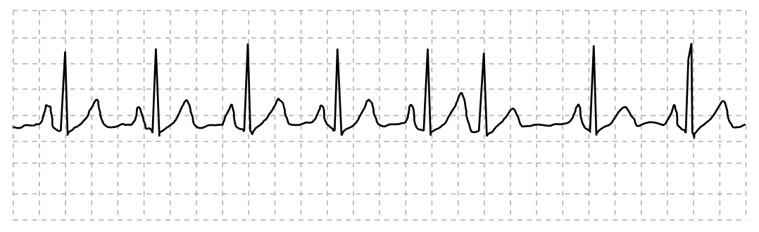
QRS duration: __________ QT duration: ____________
Ventricular rate and rhythm: _____________
Atrial rate rhythm: _____________________
PR interval: __________________________
Interpretation: ________________________
Introduction:
The electrocardiogram or ECG is a tool that is used to record a patient’s heart rate in real time. The heartbeat produces electrical signals that are recorded in this tool. It is recorded in a waveform, which has P, Q, R, S, T, and U components. The ECG is interpreted by doctors to know the condition of patients.
Explanation of Solution
The heart rate of the patient is regular with normal electrical activity of the heart. There was one PAC (premature atrial contractions), also known as atrial premature beats, at sixth position, which occurred naturally. The ECG tracing is interpreted as normal sinus rhythm.
QRS duration: The duration of the QRS complex is less than 0.12 seconds.
QT duration: It is more than 0.12 seconds.
Ventricular rate and rhythm: The ventricular rate is regular and between 60 and 100bpm. Ventricular rhythm is less than 0.12 seconds.
Atrial rate rhythm: It is regular with the heart rate up to 100 bpm and occurrence of one PAC at the 6th complex.
PR interval: Duration is less than 0.20 seconds.
Interpretation: The patient’s heart rate is between 60 and 100 bpm, so the graph represents ECG tracing of Normal Sinus Rhythm (NSR) with one PAC at the sixth complex.
QRS duration: 0.6 seconds QT duration: 0.40 seconds
Ventricular rate and rhythm: 86 bpm
Atrial rate rhythm: 86 bpm
PR interval: 0.16 seconds
Interpretation: The graph shows normal sinus rhythm with 86 bpm having one atrial premature beat at the sixth complex
Want to see more full solutions like this?
Chapter 14 Solutions
Cardiopulmonary Anatomy & Physiology


- Determine how much ATP would a cell produce when using fermentation of a 50 mM glucose solution?arrow_forwardDetermine how much ATP would a cell produce when using aerobic respiration of a 7 mM glucose solution?arrow_forwardDetermine how much ATP would a cell produce when using aerobic respiration to degrade one small protein molecule into 12 molecules of malic acid, how many ATP would that cell make? Malic acid is an intermediate in the Krebs cycle. Assume there is no other carbon source and no acetyl-CoA.arrow_forward
- Identify each of the major endocrine glandsarrow_forwardCome up with a few questions and answers for umbrella species, keystone species, redunant species, and aquatic keystone speciesarrow_forward19. On the diagram below a. Label the three pictures as: DNA; polypeptide; or RNA. b. Label the arrows as: translation or transcription/RNA processing. c. Add the following details to the diagram. Promoter region TATA box Transcription start site Transcription terminator Intron (A,B,C,D) Exons (1,2,3,4,5) Splice sites 5' cap 5' UTR (untranslated region) 3' poly A tail 3' UTR (untranslated region) Translational start (AUG) Translational stop (UGA, UAG, or UAA) N and C ends of polypeptide 0000arrow_forward
- Match the letter labels in the figure below to the terms. Some letter labels are not used. MNNNNNNIN M C B A M D F E H K G 8arrow_forwardThe diagram below illustrates a quorum sensing pathway from Staphylococcus aureus. Please answer the following questions. 1. Autoinduction is part of the quorum sensing system. Which promoter (P2 or P3) is critical for autoinduction? 2)This staphylococcus aureus grows on human wounds, causing severe infections. You would like to start a clinical trial to treat these wound infections. Please describe: a) What molecule do you recommend for the trial. Why? b) Your trial requires that Staphylococcus aureus be isolated from the wound and submitted to genome sequencing before admittance. Why? What are you testing for? 3) If a mutation arises where the Promoter P3 is constitutively active, how would that influence sensitivity to AIP? Please explain your rationale. 4) This pathway is sensitive to bacterial cell density. Describe two separate mutation that would render the pathway active independent of cell density. Briefly explain your rationale. Mutation 1 Mutation 2arrow_forwardThere is currently a H5N1 cattle outbreak in North America. According to the CDC on Feb 26*: "A multistate outbreak of HPAI A(H5N1) bird flu in dairy cows was first reported on March 25, 2024. This is the first time that these bird flu viruses had been found in cows. In the United States, since 2022, USDA has reported HPAI A(H5N1) virus detections in more than 200 mammals." List and describe two mechanisms that could lead to this H5N1 influenza strain evolving to spread in human: Mechanisms 1: Mechanisms 2: For the mutations to results in a human epidered they would need to change how the virus interacts with the human host. In the case of mutations that may promote an epidemic, provide an example for: a protein that might incur a mutation: how the mutation would change interactions with cells in the respiratory tract (name the receptor on human cells) List two phenotypic consequence from this mutation that would increase human riskarrow_forward
- You have a bacterial strain with the CMU operon: a) As shown in the image below, the cmu operon encodes a peptide (Pep1), as well as a kinase and regulator corresponding to a two-component system. The cmu operon is activated when Pep 1 is added to the growth media. Pep1 is a peptide that when added extracellularly leads to activation of the Cmu operon. Pep1 cmu-kinase cmu-regulator You also have these genetic components in other strains: b) An alternative sigma factor, with a promoter activated by the cmu-regulator, that control a series of multiple operons that together encode a transformasome (cellular machinery for transformation). c) the gene cl (a repressor). d) the promoter X, which includes a cl binding site (and in the absence of cl is active). e) the gene gp (encoding a green fluorescence protein). Using the cmu operon as a starting point, and assuming you can perform cloning to rearrange any of these genomic features, how would you use one or more of these to modify the…arrow_forwardYou have identified a new species of a Gram-positive bacteria. You would like to screen their genome for all proteins that are covalently linked to the cell wall. You have annotated the genome, so that you identified all the promoters, operons, and genes sequences within the operons. Using these features, what would you screen for to identify a set of candidates for proteins covalently linked to the bacterial cell wall.arrow_forwardBelow is a diagram from a genomic locus of a bacterial genome. Each arrow represents a coding region, and the arrowheads indicate its orientation in the genome. The numbers are randomly assigned. Draw the following features on the diagram, and explain your rationale for each feature: 10 12 合會會會會長 6 a) Expected transcriptions, based on known properties of bacterial genes and operons. How many proteins are encoded in each of the transcripts? b) Location of promoters (include rationale) c) Location of transcriptional terminators (include rationale) d) Locations of Shine-Dalgarno sequences (include rationale)arrow_forward
 Understanding Health Insurance: A Guide to Billin...Health & NutritionISBN:9781337679480Author:GREENPublisher:Cengage
 Human Physiology: From Cells to Systems (MindTap ...BiologyISBN:9781285866932Author:Lauralee SherwoodPublisher:Cengage Learning