Campbell Biology: Custom Edition
18th Edition
ISBN: 9781323717271
Author: Urry, Cain, Wasserman, Minorsky, Reece
Publisher: PEARSON C
expand_more
expand_more
format_list_bulleted
Concept explainers
Textbook Question
Chapter 12, Problem 9TYU
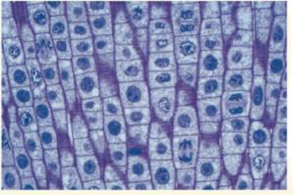
VISUAL SKILLS The light micrograph shows dividing cells near the tip of an onion root. Identify a cell in each of the following stages: prophase, prometaphase, metaphase, anaphase, and telophase. Describe the major events occurring at each stage.

Expert Solution & Answer
Trending nowThis is a popular solution!

Students have asked these similar questions
would this be considered a novel protein and if not how can I fix it so it is and can you draw the corrections please
In as much detail as possible, hand draw a
schematic diagram of the hypothalamic-pituitary-
gonad (HPG) axis in the human male. Be sure to
include all the relevant structures and hormones.
You must define all abbreviations the first time
you use them. Please include (and explain) the
feedback loops.
A negligence action was brought by a mother against a hospital on behalf of her minor daughter. It alleged that when the mother was 13 years of age, the hospital negligently transfused her with Rh-positive blood. The mother's Rh-negative blood was incompatible with and sensitized by the Rh-positive blood. The mother discovered her condition 8 years later during a routine blood screening ordered by her healthcare provider in the course of prenatal care. The resulting sensitization of the mother's blood allegedly caused damage to the fetus, resulting in physical defects and premature birth.
Did a patient relationship with the transfusing hospital exist?
Chapter 12 Solutions
Campbell Biology: Custom Edition
Ch. 12.1 - How many chromosomes are drawn in each part of...Ch. 12.1 - WHAT IF? A chicken has 78 chromosomes in its...Ch. 12.2 - How many chromosomes are shown in the illustration...Ch. 12.2 - Compare cytokinesis in animal cells and plant...Ch. 12.2 - During which stages of the cell cycle does a...Ch. 12.2 - Compare the roles of tubulin and actin during...Ch. 12.2 - A kinetochore has been compared to a coupling...Ch. 12.2 - MAKE CONNECTIONS What other functions do actin...Ch. 12.3 - In Figure 12.14, why do the nuclei resulting from...Ch. 12.3 - How does MPF allow a cell to pass the G2 phase...
Ch. 12.3 - MAKE CONNECTIONS Explain how receptor tyrosine...Ch. 12 - Differentiate between these terms: chromosome,...Ch. 12 - In which of the three phases of interphase and the...Ch. 12 - Explain the significance of the G1, G2, and M...Ch. 12 - Through a microscope, you can see a cell plate...Ch. 12 - Vinblastine is a standard chemotherapeutic drug...Ch. 12 - One difference between cancer cells and normal...Ch. 12 - The decline of MPF activity at the end of mitosis...Ch. 12 - In the cells of some organisms, mitosis occurs...Ch. 12 - Which of the following does not occur during...Ch. 12 - Cell A has half as much DNA as cells B, C, and D...Ch. 12 - The drug cytochalasin B blocks the function of...Ch. 12 - VISUAL SKILLS The light micrograph shows dividing...Ch. 12 - DRAW IT Draw one eukaryotic chromosome as it would...Ch. 12 - EVOLUTION CONNECTION The result of mitosis is that...Ch. 12 - SCIENTIFIC INQUIRY Although both ends of a...Ch. 12 - WRITE ABOUT A THEME: INFORMATION The continuity of...Ch. 12 - SYNTHESIZE YOUR KNOWLEDGE For selected answers,...
Additional Science Textbook Solutions
Find more solutions based on key concepts
Single penny tossed 20 times and counting heads and tails: Probability (prediction): _______/20 heads ________/...
Laboratory Manual For Human Anatomy & Physiology
Why do scientists think that all forms of life on earth have a common origin?
Genetics: From Genes to Genomes
Gregor Mendel never saw a gene, yet he concluded that some inherited factors were responsible for the patterns ...
Campbell Essential Biology (7th Edition)
To test your knowledge, discuss the following topics with a study partner or in writing ideally from memory. Th...
HUMAN ANATOMY
Describe the role and impact of microbes on the earth.
Microbiology Fundamentals: A Clinical Approach
Knowledge Booster
Learn more about
Need a deep-dive on the concept behind this application? Look no further. Learn more about this topic, biology and related others by exploring similar questions and additional content below.Similar questions
- 18. Watch this short youtube video about SARS CoV-2 replication. SARS-CoV-2 Life Cycle (Summer 2020) - YouTube.19. What is the name of the receptor that SARS CoV-2 uses to enter cells? Which human cells express this receptor? 20. Name a few of the proteins that the SARS CoV-2 mRNA codes for. 21. What is the role of the golgi apparatus related to SARS CoV-2arrow_forwardState the five functions of Globular Proteins, and give an example of a protein for each function.arrow_forwardDiagram of check cell under low power and high powerarrow_forward
- a couple in which the father has the a blood type and the mother has the o blood type produce an offspring with the o blood type, how does this happen? how could two functionally O parents produce an offspring that has the a blood type?arrow_forwardWhat is the opening indicated by the pointer? (leaf x.s.) stomate guard cell lenticel intercellular space none of thesearrow_forwardIdentify the indicated tissue? (stem x.s.) parenchyma collenchyma sclerenchyma ○ xylem ○ phloem none of thesearrow_forward
- Where did this structure originate from? (Salix branch root) epidermis cortex endodermis pericycle vascular cylinderarrow_forwardIdentify the indicated tissue. (Tilia stem x.s.) parenchyma collenchyma sclerenchyma xylem phloem none of thesearrow_forwardIdentify the indicated structure. (Cucurbita stem l.s.) pit lenticel stomate tendril none of thesearrow_forward
- Identify the specific cell? (Zebrina leaf peel) vessel element sieve element companion cell tracheid guard cell subsidiary cell none of thesearrow_forwardWhat type of cells flank the opening on either side? (leaf x.s.) vessel elements sieve elements companion cells tracheids guard cells none of thesearrow_forwardWhat specific cell is indicated. (Cucurbita stem I.s.) vessel element sieve element O companion cell tracheid guard cell none of thesearrow_forward
arrow_back_ios
SEE MORE QUESTIONS
arrow_forward_ios
Recommended textbooks for you
 Biology (MindTap Course List)BiologyISBN:9781337392938Author:Eldra Solomon, Charles Martin, Diana W. Martin, Linda R. BergPublisher:Cengage Learning
 Biology 2eBiologyISBN:9781947172517Author:Matthew Douglas, Jung Choi, Mary Ann ClarkPublisher:OpenStax
 Human Heredity: Principles and Issues (MindTap Co...BiologyISBN:9781305251052Author:Michael CummingsPublisher:Cengage Learning
 Biology: The Unity and Diversity of Life (MindTap...BiologyISBN:9781337408332Author:Cecie Starr, Ralph Taggart, Christine Evers, Lisa StarrPublisher:Cengage Learning
 Anatomy & PhysiologyBiologyISBN:9781938168130Author:Kelly A. Young, James A. Wise, Peter DeSaix, Dean H. Kruse, Brandon Poe, Eddie Johnson, Jody E. Johnson, Oksana Korol, J. Gordon Betts, Mark WomblePublisher:OpenStax College

Biology (MindTap Course List)
Biology
ISBN:9781337392938
Author:Eldra Solomon, Charles Martin, Diana W. Martin, Linda R. Berg
Publisher:Cengage Learning

Biology 2e
Biology
ISBN:9781947172517
Author:Matthew Douglas, Jung Choi, Mary Ann Clark
Publisher:OpenStax


Human Heredity: Principles and Issues (MindTap Co...
Biology
ISBN:9781305251052
Author:Michael Cummings
Publisher:Cengage Learning

Biology: The Unity and Diversity of Life (MindTap...
Biology
ISBN:9781337408332
Author:Cecie Starr, Ralph Taggart, Christine Evers, Lisa Starr
Publisher:Cengage Learning

Anatomy & Physiology
Biology
ISBN:9781938168130
Author:Kelly A. Young, James A. Wise, Peter DeSaix, Dean H. Kruse, Brandon Poe, Eddie Johnson, Jody E. Johnson, Oksana Korol, J. Gordon Betts, Mark Womble
Publisher:OpenStax College
The Cell Cycle and its Regulation; Author: Professor Dave Explains;https://www.youtube.com/watch?v=eqJqhA8HSJ0;License: Standard YouTube License, CC-BY
Cell Division - Mitosis and Meiosis - GCSE Biology (9-1); Author: Mr Exham Biology;https://www.youtube.com/watch?v=w7vp_uRA8kw;License: Standard YouTube License, CC-BY