Campbell Biology (11th Edition)
11th Edition
ISBN: 9780134093413
Author: Lisa A. Urry, Michael L. Cain, Steven A. Wasserman, Peter V. Minorsky, Jane B. Reece
Publisher: PEARSON
expand_more
expand_more
format_list_bulleted
Concept explainers
Videos
Textbook Question
Chapter 12, Problem 9TYU
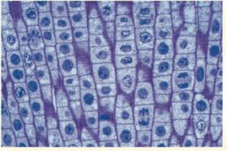
VISUAL SKILLS The light micrograph shows dividing cells near the tip of an onion root. Identify a cell in each of the following stages: prophase, prometaphase, metaphase, anaphase, and telophase. Describe the major events occurring at each stage.
Expert Solution & Answer

Trending nowThis is a popular solution!

Students have asked these similar questions
Give the terminal regression line equation and R or R2 value:
Give the x axis (name and units, if any) of the terminal line:
Give the y axis (name and units, if any) of the terminal line:
Give the first residual regression line equation and R or R2 value:
Give the x axis (name and units, if any) of the first residual line
: Give the y axis (name and units, if any) of the first residual line:
Give the second residual regression line equation and R or R2 value:
Give the x axis (name and units, if any) of the second residual line:
Give the y axis (name and units, if any) of the second residual line:
a) B1
Solution
b) B2
c)hybrid rate constant (λ1)
d)hybrid rate constant (λ2)
e) ka
f) t1/2,absorb
g) t1/2, dist
h) t1/2, elim
i)apparent central compartment volume (V1,app)
j) total AUC (short cut method)
k) apparent volume of distribution based on AUC (VAUC,app)
l)apparent clearance (CLapp)
m) absolute bioavailability of oral route (need AUCiv…
You inject morpholino oligonucleotides that inhibit the translation of follistatin, chordin, and noggin (FCN) at the 1 cell stage of a frog embryo.
What is the effect on neurulation in the resulting embryo?
Propose an experiment that would rescue an embryo injected with FCN morpholinos.
Participants will be asked to create a meme regarding a topic relevant to the department of Geography, Geomatics, and Environmental Studies.
Prompt: Using an online art style of your choice, please make a meme related to the study of Geography, Environment, or Geomatics.
Chapter 12 Solutions
Campbell Biology (11th Edition)
Ch. 12.1 - How many chromosomes are drawn in each part of...Ch. 12.1 - WHAT IF? A chicken has 78 chromosomes in its...Ch. 12.2 - How many chromosomes are shown in the illustration...Ch. 12.2 - Compare cytokinesis in animal cells and plant...Ch. 12.2 - During which stages of the cell cycle does a...Ch. 12.2 - Compare the roles of tubulin and actin during...Ch. 12.2 - A kinetochore has been compared to a coupling...Ch. 12.2 - MAKE CONNECTIONS What other functions do actin...Ch. 12.3 - In Figure 12.14, why do the nuclei resulting from...Ch. 12.3 - How does MPF allow a cell to pass the G2 phase...
Ch. 12.3 - MAKE CONNECTIONS Explain how receptor tyrosine...Ch. 12 - Differentiate between these terms: chromosome,...Ch. 12 - In which of the three phases of interphase and the...Ch. 12 - Explain the significance of the G1, G2, and M...Ch. 12 - Through a microscope, you can see a cell plate...Ch. 12 - Vinblastine is a standard chemotherapeutic drug...Ch. 12 - One difference between cancer cells and normal...Ch. 12 - The decline of MPF activity at the end of mitosis...Ch. 12 - In the cells of some organisms, mitosis occurs...Ch. 12 - Which of the following does not occur during...Ch. 12 - Cell A has half as much DNA as cells B, C, and D...Ch. 12 - The drug cytochalasin B blocks the function of...Ch. 12 - VISUAL SKILLS The light micrograph shows dividing...Ch. 12 - DRAW IT Draw one eukaryotic chromosome as it would...Ch. 12 - EVOLUTION CONNECTION The result of mitosis is that...Ch. 12 - SCIENTIFIC INQUIRY Although both ends of a...Ch. 12 - WRITE ABOUT A THEME: INFORMATION The continuity of...Ch. 12 - SYNTHESIZE YOUR KNOWLEDGE For selected answers,...
Additional Science Textbook Solutions
Find more solutions based on key concepts
Single penny tossed 20 times and counting heads and tails: Probability (prediction): _______/20 heads ________/...
Laboratory Manual For Human Anatomy & Physiology
Why do scientists think that all forms of life on earth have a common origin?
Genetics: From Genes to Genomes
Gregor Mendel never saw a gene, yet he concluded that some inherited factors were responsible for the patterns ...
Campbell Essential Biology (7th Edition)
To test your knowledge, discuss the following topics with a study partner or in writing ideally from memory. Th...
HUMAN ANATOMY
Describe the role and impact of microbes on the earth.
Microbiology Fundamentals: A Clinical Approach
Knowledge Booster


Learn more about
Need a deep-dive on the concept behind this application? Look no further. Learn more about this topic, biology and related others by exploring similar questions and additional content below.Similar questions
- Plekhg5 functions in bottle cell formation, and Shroom3 functions in neural plate closure, yet the phenotype of injecting mRNA of each into the animal pole of a fertilized egg is very similar. What is the phenotype, and why is the phenotype so similar? Is the phenotype going to be that there is a disruption of the formation of the neural tube for both of these because bottle cell formation is necessary for the neural plate to fold in forming the neural tube and Shroom3 is further needed to close the neural plate? So since both Plekhg5 and Shroom3 are used in forming the neural tube, injecting the mRNA will just lead to neural tube deformity?arrow_forwardWhat are some medical issues or health trends that may have a direct link to the idea of keeping fat out of diets?arrow_forwardwhat did charles darwin do in sciencearrow_forward
- fa How many different gametes, f₂ phenotypes and f₂ genotypes can potentially be produced from individuals of the following genotypes? 1) AaBb i) AaBB 11) AABSC- AA Bb Cc Dd EE Cal bsm nortubaarrow_forwardC MasteringHealth MasteringNu × session.healthandnutrition-mastering.pearson.com/myct/itemView?assignment ProblemID=17396416&attemptNo=1&offset=prevarrow_forward10. Your instructor will give you 2 amino acids during the activity session (video 2-7. A. First color all the polar and non-polar covalent bonds in the R groups of your 2 amino acids using the same colors as in #7. Do not color the bonds in the backbone of each amino acid. B. Next, color where all the hydrogen bonds, hydrophobic interactions and ionic bonds could occur in the R group of each amino acid. Use the same colors as in #7. Do not color the bonds in the backbone of each amino acid. C. Position the two amino acids on the page below in an orientation where the two R groups could bond together. Once you are satisfied, staple or tape the amino acids in place and label the bond that you formed between the two R groups. - Polar covalent Bond - Red - Non polar Covalent boND- yellow - Ionic BonD - PINK Hydrogen Bonn - Purple Hydrophobic interaction-green O=C-N H I. H HO H =O CH2 C-C-N HICK H HO H CH2 OH H₂N C = Oarrow_forwardFind the dental formula and enter it in the following format: I3/3 C1/1 P4/4 M2/3 = 42 (this is not the correct number, just the correct format) Please be aware: the upper jaw is intact (all teeth are present). The bottom jaw/mandible is not intact. The front teeth should include 6 total rectangular teeth (3 on each side) and 2 total large triangular teeth (1 on each side).arrow_forward12. Calculate the area of a circle which has a radius of 1200 μm. Give your answer in mm² in scientific notation with the correct number of significant figures.arrow_forwardDescribe the image quality of the B.megaterium at 1000X before adding oil? What does adding oil do to the quality of the image?arrow_forwardarrow_back_iosSEE MORE QUESTIONSarrow_forward_ios
Recommended textbooks for you
 Biology (MindTap Course List)BiologyISBN:9781337392938Author:Eldra Solomon, Charles Martin, Diana W. Martin, Linda R. BergPublisher:Cengage Learning
 Biology 2eBiologyISBN:9781947172517Author:Matthew Douglas, Jung Choi, Mary Ann ClarkPublisher:OpenStax
 Human Heredity: Principles and Issues (MindTap Co...BiologyISBN:9781305251052Author:Michael CummingsPublisher:Cengage Learning
 Biology: The Unity and Diversity of Life (MindTap...BiologyISBN:9781337408332Author:Cecie Starr, Ralph Taggart, Christine Evers, Lisa StarrPublisher:Cengage Learning
 Anatomy & PhysiologyBiologyISBN:9781938168130Author:Kelly A. Young, James A. Wise, Peter DeSaix, Dean H. Kruse, Brandon Poe, Eddie Johnson, Jody E. Johnson, Oksana Korol, J. Gordon Betts, Mark WomblePublisher:OpenStax College
Biology (MindTap Course List)
Biology
ISBN:9781337392938
Author:Eldra Solomon, Charles Martin, Diana W. Martin, Linda R. Berg
Publisher:Cengage Learning
Biology 2e
Biology
ISBN:9781947172517
Author:Matthew Douglas, Jung Choi, Mary Ann Clark
Publisher:OpenStax
Human Heredity: Principles and Issues (MindTap Co...
Biology
ISBN:9781305251052
Author:Michael Cummings
Publisher:Cengage Learning
Biology: The Unity and Diversity of Life (MindTap...
Biology
ISBN:9781337408332
Author:Cecie Starr, Ralph Taggart, Christine Evers, Lisa Starr
Publisher:Cengage Learning
Anatomy & Physiology
Biology
ISBN:9781938168130
Author:Kelly A. Young, James A. Wise, Peter DeSaix, Dean H. Kruse, Brandon Poe, Eddie Johnson, Jody E. Johnson, Oksana Korol, J. Gordon Betts, Mark Womble
Publisher:OpenStax College
The Cell Cycle and its Regulation; Author: Professor Dave Explains;https://www.youtube.com/watch?v=eqJqhA8HSJ0;License: Standard YouTube License, CC-BY
Cell Division - Mitosis and Meiosis - GCSE Biology (9-1); Author: Mr Exham Biology;https://www.youtube.com/watch?v=w7vp_uRA8kw;License: Standard YouTube License, CC-BY