Concept explainers
39-Year-Old Female with Traumatic Brain Injury
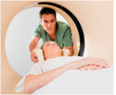
Margaret Bryans, a 39-year-old female, lost control while skiing and hit her head on a tree. She regained consciousness en route to the hospital and appeared agitated and combative. Paramedics observed that she had a right hemiparesis (muscle weakness), with a nearly complete paresis of her right upper extremity and a partial paresis of the right lower extremity. A head CT scan revealed an acute subdural hematoma and an extensive subarachnoid hemorrhage. Doctors noted that she was able to follow commands from medical personnel. However, she could only speak haltingly, using simple words.

Surgery to remove large clots from the subarachnoid space was performed immediately. Two weeks after the surgery, she showed significant improvement in her speech and motor function.
1. The adult brain consists of four main anatomical regions (see Table 12.1. pp. 458–459). Based on the observed signs in this case, which of these four brain regions is involved? What evidence did you use to determine this?
Want to see the full answer?
Check out a sample textbook solution
Chapter 12 Solutions
HUMAN ANATOMY & PHYSIOLOGY (LCPO)
- "One of the symmetry breaking events in mouse gastrulation requires the amplification of Nodal on the side of the embryo opposite to the Anterior Visceral Endoderm (AVE). Describe one way by which Nodal gets amplified in this region." My understanding of this is that there are a few ways nodal is amplified though I'm not sure if this is specifically occurs on the opposite side of the AVE. 1. pronodal cleaved by protease -> active nodal 2. Nodal -> BMP4 -> Wnt-> nodal 3. Nodal-> Nodal, Fox1 binding site 4. BMP4 on outside-> nodal Are all of these occuring opposite to AVE?arrow_forwardIf four babies are born on a given day What is the chance all four will be girls? Use genetics lawsarrow_forwardExplain each punnet square results (genotypes and probabilities)arrow_forward
- Give the terminal regression line equation and R or R2 value: Give the x axis (name and units, if any) of the terminal line: Give the y axis (name and units, if any) of the terminal line: Give the first residual regression line equation and R or R2 value: Give the x axis (name and units, if any) of the first residual line : Give the y axis (name and units, if any) of the first residual line: Give the second residual regression line equation and R or R2 value: Give the x axis (name and units, if any) of the second residual line: Give the y axis (name and units, if any) of the second residual line: a) B1 Solution b) B2 c)hybrid rate constant (λ1) d)hybrid rate constant (λ2) e) ka f) t1/2,absorb g) t1/2, dist h) t1/2, elim i)apparent central compartment volume (V1,app) j) total AUC (short cut method) k) apparent volume of distribution based on AUC (VAUC,app) l)apparent clearance (CLapp) m) absolute bioavailability of oral route (need AUCiv…arrow_forwardYou inject morpholino oligonucleotides that inhibit the translation of follistatin, chordin, and noggin (FCN) at the 1 cell stage of a frog embryo. What is the effect on neurulation in the resulting embryo? Propose an experiment that would rescue an embryo injected with FCN morpholinos.arrow_forwardParticipants will be asked to create a meme regarding a topic relevant to the department of Geography, Geomatics, and Environmental Studies. Prompt: Using an online art style of your choice, please make a meme related to the study of Geography, Environment, or Geomatics.arrow_forward
- Plekhg5 functions in bottle cell formation, and Shroom3 functions in neural plate closure, yet the phenotype of injecting mRNA of each into the animal pole of a fertilized egg is very similar. What is the phenotype, and why is the phenotype so similar? Is the phenotype going to be that there is a disruption of the formation of the neural tube for both of these because bottle cell formation is necessary for the neural plate to fold in forming the neural tube and Shroom3 is further needed to close the neural plate? So since both Plekhg5 and Shroom3 are used in forming the neural tube, injecting the mRNA will just lead to neural tube deformity?arrow_forwardWhat are some medical issues or health trends that may have a direct link to the idea of keeping fat out of diets?arrow_forwardwhat did charles darwin do in sciencearrow_forward
- Understanding Health Insurance: A Guide to Billin...Health & NutritionISBN:9781337679480Author:GREENPublisher:CengageSurgical Tech For Surgical Tech Pos CareHealth & NutritionISBN:9781337648868Author:AssociationPublisher:Cengage
 Medical Terminology for Health Professions, Spira...Health & NutritionISBN:9781305634350Author:Ann Ehrlich, Carol L. Schroeder, Laura Ehrlich, Katrina A. SchroederPublisher:Cengage Learning
 Fundamentals of Sectional Anatomy: An Imaging App...BiologyISBN:9781133960867Author:Denise L. LazoPublisher:Cengage Learning


