
Concept explainers
Answers to all problems are at the end of this book. Detailed solutions are available in the Student Solutions Manual, Study Guide, and Problems Book.
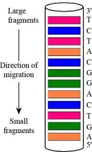
Predicting a Sanger Sequencing Pattern The oligonucleotide d-AGATGCCTGACT as subjected to sequencing by Sanger’s dideoxy method, using fluorescent-tagged dideoxynucleotides and capillary electrophoresis, essentially as shown in Figure 11.3. Draw a diagram of the gel-banding pattern within the capillary.
Interpretation: A diagram of the gel-banding pattern within the capillary is to be drawn.
Concept introduction: A laboratory technique that is used for the separation of charged molecules such as proteins, DNA and RNA on the basis of their size is known as gel electrophoresis. This technique is useful to distinguish DNA fragments of various lengths.
Answer to Problem 1P
A diagram of the gel-banding pattern within the capillary is,
Explanation of Solution
In gel electrophoresis, using dyes such as radioactive labels or fluorescent tags makes it possible to see the DNA on the gel after separation. They are going to appear on the gel as bands. Therefore, the fluorescently labeled dideoxynucleotides result in the formation of the gel banding pattern. The labeled dideoxynucleotides are then added to the growing chain of DNA and capillary electrophoresis is applied to the resulting fragments.
The given oligonucleotide is d-AGATGCCTGACT that was subjected to sequencing by Sanger’s dideoxy method. In gel-banding pattern within the capillary, the top of the column consists of larger fragments and the bottom of the column has smaller fragments. The
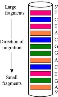
Figure 1
Want to see more full solutions like this?
Chapter 11 Solutions
Biochemistry
- Answers to all problems are at the end of this book. Detailed solutions are available in the Student Solutions Manual, Study Guide, and Problems Book. Designing Primers for PCR Amplification of a DNA Sequence Given the following short DNA duplex of sequence (53)ATGCCGTAGTCGATCATTACGATAGCATAGCACAGGGATCCA- CATGCACACACATGACATAGGACAGATAGCAT what oligonucleotide primers (17-mers) would be required for PCR amplification of this duplex?arrow_forwardAnswers to all problems are at the end of this book. Detailed solutions are available in the Student Solutions Manual, Study Guide, and Problems Book. Deducing DNA Sequence from Sanger Sequencing Results The output of an automated DNA sequence determination by the Sanger dideoxy chain termination method, performed as illustrated in Figure 11.3, is disp1ayed at right. What is the sequence of the original oligonucleotide?arrow_forwardAnswers to all problems are at the end of this book. Detailed solutions are available in the Student Solutions Manual, Study Guide, and Problems Book. CRISPR/Cas9: Design of a gRNA to Target the Human PVALB Gene The human PVALB gene, which encodes the Ca2+-binding protein parvalbumin, can be Targeted by CRISPR/Cas9, at the protospacer sequence - ATGCAGGAGGGTGGCGAGAGGGGCCGAGAT- followed by a -TGG-PAM trinucleotide. Give the sequence of the spacer region of a gRNA that will target the complementary DNA strand at this site. Include at the 3'-end of your gRNA sequence a region that will form a stem-loop structure with a 5'-AGCAUAGCUGUAAAAC- sequence downstream in the gRNA to create the dsRNA-binding site for Cas9.arrow_forward
- Answers to all problems are at the end of this book. Detailed solutions are available in the Student Solutions Manual, Study Guide, and Problems Book. Preparing cDNA Libraries from Different Cells Describe an experimental protocol for the preparation of to cDNA libraries, one from anaerobically grown yeast cells and the second from aerobically grown yeast cell.arrow_forwardAnswers to all problems are at the end of this book. Detailed solutions are available in the Student Solutions Manual, Study Guide, and Problems Book. Identifying DNA Structural and Functional Elements from Nucleotide Sequence Information Listed below are four DNA sequences. Which one contains a type-II restriction endonuclease (six-cutter) hexanucleotide site?. Which one is likely to form a cruciform structure? Which one is likely to be found in Z-DNA? Which one represents the 5'-end of a tRNA gene? Which one is most likely to be found in a triplex DNA structure? a. CGCGCGCCGCGCACGCGCTCGCGCGCCGC b. GAACGTCGTATTCCCGTACGACGTTC c. CAGGTCTCTCTCTCTCTCTCTC d. TGGTGCGAATTCTGTGGAT e. ATCGGAATTCATCGarrow_forwardAnswers to all problems are at the end of this book. Detailed solutions are available in the Student Solutions Manual, Study Guide, and Problems Book. The Sequence Relationship Between an Antisense RNA Strand and Its Template DNA Strand The DNA strand that is complementary to the template strand copied by RNA polymerase during transcription has a nucleotide sequence identical to that of the RNA being synthesized (except T residues are found in the DNA strand at sites where U residues occur in the RNA). An RNA transcribed from this nontem-plate DNA strand would be complementary to the mRNA synthesized by RNA polymerase. Such an RNA is called antisense RNA because its base sequence is complementary to the “sense mRNA. One strategy to thwart the deleterious effects of genes activated in disease slates (such as cancer) is to generate antisense RNAs in affected cells. These antisense RNAs would form double-stranded hybrids with mRNAs transcribed from the activated genes and prevent their translation into protein. Suppose transcription of a cancer-activated gene yielded an mRNA whose sequence included the segment 5’-UACGUCUAAGCUGA. What is the corresponding nucleotide sequence (5’ The template strand in a DNA duplex that might be introduced into these cells so that an untisense RNA could be transcribed from it?arrow_forward
- Answers to all problems are at the end of this book. Detailed solutions are available in the Student Solutions Manual, Study Guide, and Problems Book. B- and Z-DNA Supercoiling Parameters Suppose one double helical turn of a superhelical DNA molecule changes conformation from B- to Z-form. What are the changes in L, W, and T? Why do you suppose the transition of DNA from B- to Z-form is favored by negative supercoiling?arrow_forwardAnswers to all problems are at the end of this book. Detailed solutions are available in the Student Solutions Manual, Study Guide, and Problems Book. B-and Z-DNA Helical Parameters II If 80% of the base pairs in a duplex DNA molecule (12.5 kbp) are in the B-conformation and 20% are in the Z-conformation, what is the length of the molecule?arrow_forwardAnswers to all problems are at the end of this book. Detailed solutions are available in the Student Solutions Manual, Study Guide, and Problems Book. DNA Supercoiling Parameters A “relaxed,� circular, double-stranded DNA molecule (1600 bp) is in a solution where conditions favor 10 bp per turn. What is the value of L0 for this DNA molecule? Suppose DNA gyrase introduces 12 negative supercoils into this molecule. What are the values of L, W, and T now? What is the superhelical density, ?arrow_forward
- Answers to all problems are at the end of this book. Detailed solutions are available in the Student Solutions Manual, Study Guide, and Problems Book. The Base Sequence in the Two Polynucleotide Chairs of a DNA Double Helix Is Complementary Adhering to the convention of writing nucleotide sequences in the 5' 3' direction, what is the nucleotide sequence of the DNA strand that is complementary to d-ATCGCAACTGTCACTA?arrow_forwardAnswers to all problems are at the end of this book. Detailed solutions are available in the Student Solutions Manual, Study Guide, and Problems Book. Structural complementarity is the key to molecular recognition, a lesson learned in Chapter 1. The principle of structural complementarity is relevant to answering problems 5, 6, 7,11, 12, and 19. The quintessential example of structural complementarity in all of biology is the DNA double helix. What features of the DNA double helix exemplify structural complementarity?arrow_forwardAnswers to all problems are at the end of this book. Detailed solutions are available in the Student Solutions Manual, Study Guide, and Problems Book. An Estimation of Minimal Genome Size for a Living Cell Studies of existing cells to determine the minimum number of genes for a living cell have suggested that 206 genes are sufficient. If the ratio of protein-coding genes to non-protein-coding genes is the same in this minimal organism as the genes of Mycoplasma genitulium, how many proteins are represented in these 206 genes. How many base pairs would be required to form the genome of this minimal organism if the genes are the same size as M genilalium genes? (Section 1.5)arrow_forward
- BiochemistryBiochemistryISBN:9781305577206Author:Reginald H. Garrett, Charles M. GrishamPublisher:Cengage Learning