Videos
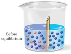
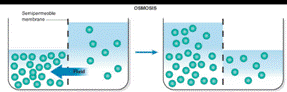
Assume that two liquids are separated by a semipermeable membrane, with pure solvent on the right side and a solution of a solute on the left side. Make a drawing that shows the situation after equilibrium is reached.
Interpretation:
The situation after equilibrium is reached when two liquids are separated by a semipermeable membrane has to be drawn.
Concept introduction:
Colligative properties such as vapor pressure, freezing point, and boiling point are affected by the presence of solute particles in a solution.
Osmosis is the passage of substances in true solution through a semipermeable membrane.
Particles such as starches and protein molecules are too large to pass the membrane.
The restriction of passage of large particles causes the smaller particles to pass more rapidly in the direction of higher concentrations, producing an osmotic pressure in confined liquids.
Explanation of Solution
Osmosis occurs when solution of different concentration are separated by a semipermeable membrane that allows solvent molecules to pass but blocks the passage of solute ions and molecules. Solvent flows from the more dilute side to the more concentrated side until sufficient osmotic pressure builds up and stops the flows. An effect similar to osmosis occurs when membranes of large pore size are used.
The situation after equilibrium is reached when two liquids are separated by a semipermeable membrane is drawn as
Want to see more full solutions like this?
Chapter 9 Solutions
Fundamentals of General, Organic, and Biological Chemistry (8th Edition)


- 10. Which one of the following compounds is the major organic product of the series of reactions shown here? Ph A OH Ph B CO₂Et Br -H Ν ΚΑ CO₂Et 1. NaOEt 1. NaOH, H₂O 2. H3O+ 2. PhCH2CH2Br 3. heat NH2 Ph OH NH2 0 OH Ph OH NH2 Ph D NH2 E OHarrow_forward1. What is the isoelectric point of glutamic acid? (Glutamic acid has pKa1 2.10, pKa2 4.07, pKaз 9.47) A) pH 2.1 D) pH 6.8 B) pH 3.1 C) pH 4.1 E) pH 9.5 HO NH2 Glutamic acid (shown without charges) OHarrow_forwardBr Mg, ether 1. HCHO (formaldehyde) 2. H+, H₂O PCC 1. NH3, HCN ? (pyridinium chlorochromate) 2. H2O, HCI 11. Which one of the following compounds is the major organic product of the series of reactions shown above? Ph. Ph. OH NH2₂ A Ph. Ή NH2 B OH Ph Η Ph OH NH2 NH2₂ NH₂ C D Earrow_forward
- B A 6. Which ONE of the labeled bonds in the tripeptide on the right is a peptide bond: H₂N N 'N' OH C H A, B, C, D or E? HN E OHarrow_forwardQuestions 8-9 are 0.4 points each. The next two questions relate to the peptide whose structure is shown here. To answer these questions, you should look at a table of H2N/.. amino acid structures. You don't have to memorize the structures of the amino acids. IZ 8. What is the N-terminal amino acid of this peptide? A) proline B) aspartic acid C) threonine 9. What is the C-terminal amino acid of this peptide? A) proline B) aspartic acid C) threonine N OH D) valine E) leucine D) valine E) leucine NH "OH OHarrow_forward7. What is the correct name of the following tripeptide? A) Ile-Met-Ser B) Leu-Cys-Thr C) Val-Cys-Ser D) Ser-Cys-Leu E) Leu-Cys-Ser H₂N!!!!! N H ΖΙ .SH SF H IN OH OHarrow_forward
- Please draw out the following metabolic pathways: (Metabolic Map) Mitochondrion: TCA Cycle & GNG, Electron Transport, ATP Synthase, Lipolysis, Shuttle Systems Cytoplasm: Glycolysis & GNG, PPP (Pentose Phosphate Pathway), Glycogen, Lipogenesis, Transporters and Amino Acids Control: Cori/ Glc-Ala cycles, Insulin/Glucagon Reg, Local/Long Distance Regulation, Pools Used Correctlyarrow_forwardPlease help provide me an insight of what to draw for the following metabolic pathways: (Metabolic Map) Mitochondrion: TCA Cycle & GNG, Electron Transport, ATP Synthase, Lipolysis, Shuttle Systems Cytoplasm: Glycolysis & GNG, PPP (Pentose Phosphate Pathway), Glycogen, Lipogenesis, Transporters and Amino Acids Control: Cori/ Glc-Ala cycles, Insulin/Glucagon Reg, Local/Long Distance Regulation, Pools Used Correctlyarrow_forwardwrite ionization equilibriumarrow_forward
- write the ionization equilibrium for cysteine and calculate the piarrow_forwardplease answerarrow_forwardf. The genetic code is given below, along with a short strand of template DNA. Write the protein segment that would form from this DNA. 5'-A-T-G-G-C-T-A-G-G-T-A-A-C-C-T-G-C-A-T-T-A-G-3' Table 4.5 The genetic code First Position Second Position (5' end) U C A G Third Position (3' end) Phe Ser Tyr Cys U Phe Ser Tyr Cys Leu Ser Stop Stop Leu Ser Stop Trp UCAG Leu Pro His Arg His Arg C Leu Pro Gln Arg Pro Leu Gin Arg Pro Leu Ser Asn Thr lle Ser Asn Thr lle Arg A Thr Lys UCAG UCAC G lle Arg Thr Lys Met Gly Asp Ala Val Gly Asp Ala Val Gly G Glu Ala UCAC Val Gly Glu Ala Val Note: This table identifies the amino acid encoded by each triplet. For example, the codon 5'-AUG-3' on mRNA specifies methionine, whereas CAU specifies histidine. UAA, UAG, and UGA are termination signals. AUG is part of the initiation signal, in addition to coding for internal methionine residues. Table 4.5 Biochemistry, Seventh Edition 2012 W. H. Freeman and Company B eviation: does it play abbreviation:arrow_forward

 Principles Of Radiographic Imaging: An Art And A ...Health & NutritionISBN:9781337711067Author:Richard R. Carlton, Arlene M. Adler, Vesna BalacPublisher:Cengage Learning
 Concepts of BiologyBiologyISBN:9781938168116Author:Samantha Fowler, Rebecca Roush, James WisePublisher:OpenStax College
 Human Biology (MindTap Course List)BiologyISBN:9781305112100Author:Cecie Starr, Beverly McMillanPublisher:Cengage Learning