CAMPBELL BIOLOGY IN FOCUS-W/MASTR.BIO.
3rd Edition
ISBN: 9780134875040
Author: Urry
Publisher: PEARSON
expand_more
expand_more
format_list_bulleted
Textbook Question
Chapter 9, Problem 7TYU
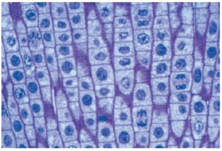
The light micrograph shows dividing cells near the tip of an onion root. Identify a cell in each of the following stages: prophase, prometaphase, metaphase, anaphase, and telophase. Describe the major events occurring at each stage.

Expert Solution & Answer
Want to see the full answer?
Check out a sample textbook solution
Students have asked these similar questions
would this be considered a novel protein and if not how can I fix it so it is and can you draw the corrections please
In as much detail as possible, hand draw a
schematic diagram of the hypothalamic-pituitary-
gonad (HPG) axis in the human male. Be sure to
include all the relevant structures and hormones.
You must define all abbreviations the first time
you use them. Please include (and explain) the
feedback loops.
A negligence action was brought by a mother against a hospital on behalf of her minor daughter. It alleged that when the mother was 13 years of age, the hospital negligently transfused her with Rh-positive blood. The mother's Rh-negative blood was incompatible with and sensitized by the Rh-positive blood. The mother discovered her condition 8 years later during a routine blood screening ordered by her healthcare provider in the course of prenatal care. The resulting sensitization of the mother's blood allegedly caused damage to the fetus, resulting in physical defects and premature birth.
Did a patient relationship with the transfusing hospital exist?
Chapter 9 Solutions
CAMPBELL BIOLOGY IN FOCUS-W/MASTR.BIO.
Ch. 9.1 - How many chromosomes are drawn in each part of...Ch. 9.1 - WHAT IF? A chicken has 78 chromosomes in its...Ch. 9.2 - How many chromosomes are shown in the drawing in...Ch. 9.2 - Compare cytokinesis in animal cells and plant...Ch. 9.2 - Prob. 3CCCh. 9.2 - Compare the roles of tubulin and actin during...Ch. 9.3 - Prob. 1CCCh. 9.3 - Prob. 2CCCh. 9.3 - Compare and contrast a benign tumor and a...Ch. 9.3 - Prob. 4CC
Ch. 9 - Through a microscope, you can see a cell plate...Ch. 9 - In the cells of some organisms, mitosis occurs...Ch. 9 - Which of the following does not occur during...Ch. 9 - Cell A has half as much DNA as cells B, C, and...Ch. 9 - The drug cytochalasin B blocks the function of...Ch. 9 - DRAW IT Draw one eukaryotic chromosome as it would...Ch. 9 - The light micrograph shows dividing cells near the...Ch. 9 - SCIENTIFIC INQUIRY Although both ends of a...Ch. 9 - FOCUS ON EVOLUTION The result of mitosis is that...Ch. 9 - FOCUS ON INFORMATION The continuity of life is...Ch. 9 - SYNTHESIZE YOUR KNOWLEDGE Shown here are two He La...
Additional Science Textbook Solutions
Find more solutions based on key concepts
An aluminum calorimeter with a mass of 100 g contains 250 g of water. The calorimeter and water are in thermal ...
Physics for Scientists and Engineers
Why do scientists think that all forms of life on earth have a common origin?
Genetics: From Genes to Genomes
Gregor Mendel never saw a gene, yet he concluded that some inherited factors were responsible for the patterns ...
Campbell Essential Biology (7th Edition)
To test your knowledge, discuss the following topics with a study partner or in writing ideally from memory. Th...
HUMAN ANATOMY
An obese 55-year-old woman consults her physician about minor chest pains during exercise. Explain the physicia...
Biology: Life on Earth with Physiology (11th Edition)
Knowledge Booster
Learn more about
Need a deep-dive on the concept behind this application? Look no further. Learn more about this topic, biology and related others by exploring similar questions and additional content below.Similar questions
- 18. Watch this short youtube video about SARS CoV-2 replication. SARS-CoV-2 Life Cycle (Summer 2020) - YouTube.19. What is the name of the receptor that SARS CoV-2 uses to enter cells? Which human cells express this receptor? 20. Name a few of the proteins that the SARS CoV-2 mRNA codes for. 21. What is the role of the golgi apparatus related to SARS CoV-2arrow_forwardState the five functions of Globular Proteins, and give an example of a protein for each function.arrow_forwardDiagram of check cell under low power and high powerarrow_forward
- a couple in which the father has the a blood type and the mother has the o blood type produce an offspring with the o blood type, how does this happen? how could two functionally O parents produce an offspring that has the a blood type?arrow_forwardWhat is the opening indicated by the pointer? (leaf x.s.) stomate guard cell lenticel intercellular space none of thesearrow_forwardIdentify the indicated tissue? (stem x.s.) parenchyma collenchyma sclerenchyma ○ xylem ○ phloem none of thesearrow_forward
- Where did this structure originate from? (Salix branch root) epidermis cortex endodermis pericycle vascular cylinderarrow_forwardIdentify the indicated tissue. (Tilia stem x.s.) parenchyma collenchyma sclerenchyma xylem phloem none of thesearrow_forwardIdentify the indicated structure. (Cucurbita stem l.s.) pit lenticel stomate tendril none of thesearrow_forward
- Identify the specific cell? (Zebrina leaf peel) vessel element sieve element companion cell tracheid guard cell subsidiary cell none of thesearrow_forwardWhat type of cells flank the opening on either side? (leaf x.s.) vessel elements sieve elements companion cells tracheids guard cells none of thesearrow_forwardWhat specific cell is indicated. (Cucurbita stem I.s.) vessel element sieve element O companion cell tracheid guard cell none of thesearrow_forward
arrow_back_ios
SEE MORE QUESTIONS
arrow_forward_ios
Recommended textbooks for you
 Biology 2eBiologyISBN:9781947172517Author:Matthew Douglas, Jung Choi, Mary Ann ClarkPublisher:OpenStax
 Human Biology (MindTap Course List)BiologyISBN:9781305112100Author:Cecie Starr, Beverly McMillanPublisher:Cengage Learning
 Biology (MindTap Course List)BiologyISBN:9781337392938Author:Eldra Solomon, Charles Martin, Diana W. Martin, Linda R. BergPublisher:Cengage Learning
 Concepts of BiologyBiologyISBN:9781938168116Author:Samantha Fowler, Rebecca Roush, James WisePublisher:OpenStax College
 Human Heredity: Principles and Issues (MindTap Co...BiologyISBN:9781305251052Author:Michael CummingsPublisher:Cengage Learning

Biology 2e
Biology
ISBN:9781947172517
Author:Matthew Douglas, Jung Choi, Mary Ann Clark
Publisher:OpenStax

Human Biology (MindTap Course List)
Biology
ISBN:9781305112100
Author:Cecie Starr, Beverly McMillan
Publisher:Cengage Learning

Biology (MindTap Course List)
Biology
ISBN:9781337392938
Author:Eldra Solomon, Charles Martin, Diana W. Martin, Linda R. Berg
Publisher:Cengage Learning

Concepts of Biology
Biology
ISBN:9781938168116
Author:Samantha Fowler, Rebecca Roush, James Wise
Publisher:OpenStax College


Human Heredity: Principles and Issues (MindTap Co...
Biology
ISBN:9781305251052
Author:Michael Cummings
Publisher:Cengage Learning
cell division of meiosis and mitosis; Author: Stated Clearly;https://www.youtube.com/watch?v=A-mFPZLLbHI;License: Standard youtube license