To describe: The signs of inflammation that came in the given statement.
Introduction: An inflammation is the sequence of events in response to the infections that would limit the effects of injury or any agent in the body. It is the biological response against the harmful stimuli to eliminate the initial cause of the healthy tissue injury or damage. Its main function is to act as a defense mechanism of the body to remove the injurious agents from the body to prevent infections.
Explanation of Solution
Given statement: While jogging in the surf, a person’s toes land on a jelly fish. The bottom of the toes becomes swollen, red, and warm to touch.
In the given case, the sting of the jellyfish causes inflammation that accompanies the visible signs such as redness, swelling, warmth, and pain. The inflammation begins through a signal provided by the damage caused by the sting. The basic steps involved in the inflammation are caused by the sting of jelly fish as follows:
Basic steps involved:
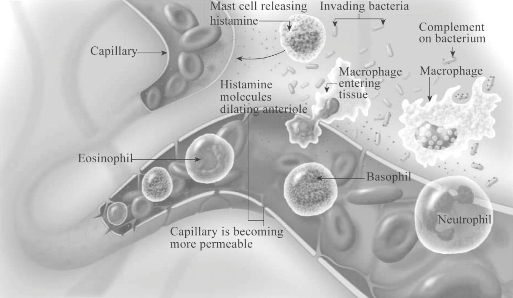
- 1. Release of various chemicals: Numerous chemicals such as histamine, interleukins, tumor necrosis factors (TNFs), chemotactic factors, leukotriene, and prostaglandins are released by the damaged cells of injured tissues basophils, mast cells, macrophages, dendritic cells, and infectious organisms to promote inflammation.
- 2. Vascular changes: The released chemical causes many local effects on the blood vessels such as:
- Increased vascular permeability
- Arteriolar vasodilation
- Leukocyte adhesion
- 3. Leukocyte recruitment:The movement of the leukocyte through the blood at the site of infection occurs by the following processes:
- a) Margination: The cell adhesion molecules (CAM) present on the surface of the leukocytes (first neutrophil and then macrophages) adhere to the endothelial capillaries present within the injured tissue.
- b) Diapedesis: Here, the cells migrate to the site of infection by exiting the blood through “squeezing out” between the vessel walls.
- c)
Chemotaxis : Chemotaxis is the movement of cells along a chemical gradient formed due to the chemicals release by the damaged cells, dead cells, or invading pathogens. Cytokines and granulocyte-macrophage colony-stimulating factor (GM-CSF) are released to attract more macrophages to the site of infection. IL-1 released by macrophages induces fever in the body. - d) Delivery of plasma proteins: Several plasma proteins such as immunoglobulin’s complement, clotting proteins, and kinins are brought into the infection site that cause local effects such as increased vascular permeability, pain, heat, and swelling on the infected site.
- 4. Tissue repair and healing:Immune cells and their secretions process the pathogen and the newly formed lymph carries the pathogen with the unwanted dead cells and cell debris from the interstitial space of the tissue. The exudate (fluid and cellular/protein mix) is passed through the lymph nodes for monitoring, transported liver, and then, removed from the body.
- 1. Redness: Inflammation increases the vascular permeability which causes the flow of a large amount of blood toward the site of infection. Due to the increased blood flow, there will be redness at the site of infection.
- 2. Heat: The heat will be generated at the site of infection due to the increased blood flow as well as the increased
metabolic activity of cells within the area. - 3. Swelling: As a result of inflammation, a large amount of fluid is lost from capillaries into the interstitial space that causes swelling around the injured site.
- 4. Pain: Pain occurs when the pain receptors are stimulated due to the compression from the accumulation of interstitial fluid and by the irritation of the released microbial substances, kinins, or prostaglandins.
- 5. Loss of function: Loss of function occurs due to the pain and swelling.
The cardinal signs of inflammation are as follows:
Thus, several immune factors produced as a result of inflammation neutralize the toxin from the jellyfish.
Pictorial representation: Fig.1 shows the steps in the inflammatory response followed by bacterial invasion and activation of immune cells at the site of injury.

Fig.1: Basic steps in inflammatory response
Want to see more full solutions like this?
Chapter 9 Solutions
Bundle: Human Biology, Loose-leaf Version, 11th + MindTap Biology, 1 term (6 months) Printed Access Card
- Describe the principle of homeostasis.arrow_forwardExplain how the hormones of the glands listed below travel around the body to target organs and tissues : Pituitary gland Hypothalamus Thyroid Parathyroid Adrenal Pineal Pancreas(islets of langerhans) Gonads (testes and ovaries) Placentaarrow_forwardWhat are the functions of the hormones produced in the glands listed below: Pituitary gland Hypothalamus Thyroid Parathyroid Adrenal Pineal Pancreas(islets of langerhans) Gonads (testes and ovaries) Placentaarrow_forward
- Describe the hormones produced in the glands listed below: Pituitary gland Hypothalamus Thyroid Parathyroid Adrenal Pineal Pancreas(islets of langerhans) Gonads (testes and ovaries) Placentaarrow_forwardPlease help me calculate drug dosage from the following information: Patient weight: 35 pounds, so 15.9 kilograms (got this by dividing 35 pounds by 2.2 kilograms) Drug dose: 0.05mg/kg Drug concentration: 2mg/mLarrow_forwardA 25-year-old woman presents to the emergency department with a 2-day history of fever, chills, severe headache, and confusion. She recently returned from a trip to sub-Saharan Africa, where she did not take malaria prophylaxis. On examination, she is febrile (39.8°C/103.6°F) and hypotensive. Laboratory studies reveal hemoglobin of 8.0 g/dL, platelet count of 50,000/μL, and evidence of hemoglobinuria. A peripheral blood smear shows ring forms and banana-shaped gametocytes. Which of the following Plasmodium species is most likely responsible for her severe symptoms? A. Plasmodium vivax B. Plasmodium ovale C. Plasmodium malariae D. Plasmodium falciparumarrow_forward
- please fill in missing parts , thank youarrow_forwardplease draw in the answers, thank youarrow_forwarda. On this first grid, assume that the DNA and RNA templates are read left to right. DNA DNA mRNA codon tRNA anticodon polypeptide _strand strand C с A T G A U G C A TRP b. Now do this AGAIN assuming that the DNA and RNA templates are read right to left. DNA DNA strand strand C mRNA codon tRNA anticodon polypeptide 0 A T G A U G с A TRParrow_forward
 Human Biology (MindTap Course List)BiologyISBN:9781305112100Author:Cecie Starr, Beverly McMillanPublisher:Cengage Learning
 Human Heredity: Principles and Issues (MindTap Co...BiologyISBN:9781305251052Author:Michael CummingsPublisher:Cengage Learning
 Biology (MindTap Course List)BiologyISBN:9781337392938Author:Eldra Solomon, Charles Martin, Diana W. Martin, Linda R. BergPublisher:Cengage Learning
 Human Physiology: From Cells to Systems (MindTap ...BiologyISBN:9781285866932Author:Lauralee SherwoodPublisher:Cengage Learning
 Anatomy & PhysiologyBiologyISBN:9781938168130Author:Kelly A. Young, James A. Wise, Peter DeSaix, Dean H. Kruse, Brandon Poe, Eddie Johnson, Jody E. Johnson, Oksana Korol, J. Gordon Betts, Mark WomblePublisher:OpenStax College
 Concepts of BiologyBiologyISBN:9781938168116Author:Samantha Fowler, Rebecca Roush, James WisePublisher:OpenStax College


