Concept explainers
Boils in the Locker Room

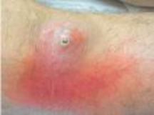
For several weeks, faculty, students, and staff at Rayburn High School have been dealing with an epidemic of unsightly, painful boils and skin infections. Over 40 athletes have been too sore to play sports, and 13 students have been hospitalized. Nurses take clinical samples of the drainage of the infections from hospitalized students, and medical laboratory scientists culture the samples. Isolated bacterial colonies are circular, convex, and golden. The bacterium is Gram-positive, mesophilic, and facultatively halophilic and can grow with or without oxygen. The cells are spherical and remain attached in clusters.
- 1. What color are the
Gram-stained cells? - 2. What does the term “facultatively halophilic” mean?
- 3. What is the scientific description of the bacterium's oxygen requirement?
- 4. If the bacterium divides every 30 minutes under laboratory conditions, how many cells would there be in a colony after 24 hours?
Want to see the full answer?
Check out a sample textbook solution
Chapter 6 Solutions
Microbiology with Diseases by Body System & Modified MasteringMicrobiology with Pearson eText -- ValuePack Access Card -- for Microbiology with Diseases by Body System Package
- I completed the chart, and I'm supposed to figure out the unknown bacteria, there are 12 unknown culture, out of which of them resemble my chart the most? (My chart is the one in pink with the information filled out)arrow_forwarddescribe the shape of the bacterial cells for each of the two yogurt bacteria. Be sure to use the terms for bacteria shapes that you learned in the Survey of the Microbial World lab!arrow_forwardTom streak plated an Unknown bacterium on Monday. On Wednesday, her plate showed a continuous streak of growth in two colors, but no distinct single colonies. List two possible reasons for these observations and why they may have affected the streak.arrow_forward
- These is another lab that we did I have finish the front but I don't understand page 24 number 4 it is asking about the Bacterial Morphology by simple stain of the Serratia Marcescens.arrow_forwardA farmer working on a piece of machinery gets his shirtsleeve caught in a moving piece of the equipment. His shirt is sliced, and a sharp blade covered in mud slices through his upper arm. He attempts to control the bleeding and immediately seeks medical attention. After 3 days, he develops a fever and his arm becomes extremely swollen and painful. Pulling back the bandages, he finds that the wound has become blackened and is leaking bloody fluid. Microscopic analysis of the fluid reveals the presence of gram-positive bacilli. a. Discuss what condition the patient is suffering from and the likely causative agent of this infection. b. Explain how the patient contracted this pathogenic microbe and what virulence factors contributed to the pathogenesis seen at the wound site. c. In addition to antibiotics, the physician prescribes hyperbaric therapy. Describe what this treatment involves and how it could be therapeutic to this patient.arrow_forwardWhat type shape is this bacteria exhibiting? streptococci spirilla staphylococci staphlobacilliarrow_forward
- The peptidoglycan layer of Gram-positive bacteria is exposed to the extracellular environment. True Falsearrow_forwardWhat is the correct genus and species of your unknown Gram (+) bacterium?arrow_forwardAbigail did not dry the top of her agar plate and stored it top up. Now she wants to count colonies but there is a smear over the entire plate. She was working with E. coli. Why does she have a smear?arrow_forward
- Which of the following diseases is NOT associated with bacteria that form endospores? tetanus anthrax toxic shock syndrome botulism gangrenearrow_forwardI'm having trouble identifying the type of bacteria I have cultured. I know that it is gram-negative and cocci in shape, it grows wonderfully on the MSA plate but did not grow so well on the EMB plate, and it is non-motile. It has a yellow color but I'm just not sure what it is. It was obtained from a public area. Any ideas?arrow_forwardTwo patients, with identical medical histories are in adjacent hospital beds. Each is infected with Streptococcus pneumoniae. Patient A's infection progresses to pneumonia while Patient B's infection resolves quickly. The microbial stain of the lung fluid from Patient A indicated the bacterial cells had a capsule, while Patient B's lung fluid stain did not. Why did the capsule affect the course of the infection in Patient A?arrow_forward
- Basic Clinical Lab Competencies for Respiratory C...NursingISBN:9781285244662Author:WhitePublisher:Cengage


