
Concept explainers
Videos
To analyze:
The inference that can be deciphered from the graph showing the relative fluidity of cell membranes in different species and whether the trend shown is apparent.
Given:
Researchers conducted an experiment to comparatively study the fluidity and composition of cell membranes. They maintained arctic sculpin at 0°C, a group of goldfish at 5°C, other group of goldfish at 25°C. They also maintained the desert pupfish at 34°C, and rats at normal temperature of 21°C. After keeping them for several days, the neuronal cells of all the animals were taken and membranes were isolated.
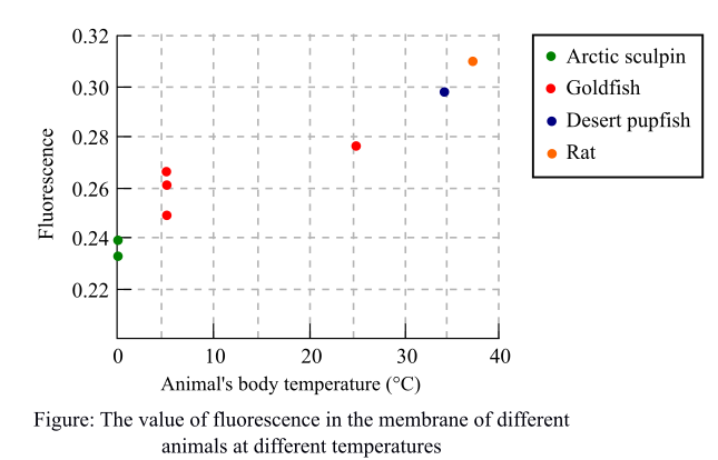
A fluorescent molecule was added to each of the extracted membranes and kept for some time at 20°C. Researchers then measured fluorescence and a graph was plotted by them. The graph depicted fluorescence against the body temperature of each of the animals.
The following graph shows the fluorescence of each animal depicted as colorful points at different temperatures. The more the value of fluorescence, the less will be the movement of molecules depicting a less fluid membrane.
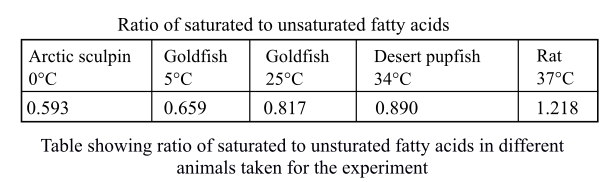
The following table was drawn by the researchers showing the ratio of saturated-to-unsaturated fatty acids in the phospholipid phosphatidyl choline for different animals taken into consideration.
Introduction:
The membrane is fluid in nature and is responsible for the transport of various molecules in and outside the cell. The fluidity of the membrane depends on the temperature and unsaturation. The low temperature leads to increase in fluidity to cope up the temperature. The saturated fatty acids are those fatty acids which do not have any double bond and have no scope of addition of any molecule. They can only substitute the molecules. The unsaturated fatty acids are those which have one or more double or triple bonds that increase the fluidity of the membrane. The species that survive in lower temperature have more amounts of unsaturated fatty acids so that they can avoid being frozen and remain fluid for transportation of the molecules.
Explanation of Solution
The data in the graph shows the value of fluorescence in different species at different temperature. It was observed that when the temperature of the animal’s body is low as in the case of arctic sculpin, the value of fluorescence is also low. There is an inverse relation between the movement of molecules and value of fluorescence. The low fluorescence depicts the high movement of molecules in the artic sculpin showing that the membrane has greater fluidity. The greater fluidity of the membrane helps these organisms to withstand such low temperature. In contrast, the fluidity of membranes of rats is low as the fluorescence value shown in the graph is high. This indicates that fluidity of membrane is not as high as in the species of low temperature.
The data of membrane fluidity depends on the temperature of the animal and is apparent as the fluidity of the membrane will change according to the change in the temperature.
Thus, it was concluded that the post translational modifications are important for the proper protein functioning and deoxyribonucleic acid (DNA) of the protein decides whether the modification should occur or not. The trend shown in the graph is apparent.
Want to see more full solutions like this?
Chapter 6 Solutions
EBK LIFE: THE SCIENCE OF BIOLOGY


- 22. Which of the following mutant proteins is expected to have a dominant negative effect when over- expressed in normal cells? a. mutant PI3-kinase that lacks the SH2 domain but retains the kinase function b. mutant Grb2 protein that cannot bind to RTK c. mutant RTK that lacks the extracellular domain d. mutant PDK that has the PH domain but lost the kinase function e. all of the abovearrow_forwardWhat is the label ?arrow_forwardCan you described the image? Can you explain the question as well their answer and how to get to an answer to an problem like this?arrow_forward
- Describe the principle of homeostasis.arrow_forwardExplain how the hormones of the glands listed below travel around the body to target organs and tissues : Pituitary gland Hypothalamus Thyroid Parathyroid Adrenal Pineal Pancreas(islets of langerhans) Gonads (testes and ovaries) Placentaarrow_forwardWhat are the functions of the hormones produced in the glands listed below: Pituitary gland Hypothalamus Thyroid Parathyroid Adrenal Pineal Pancreas(islets of langerhans) Gonads (testes and ovaries) Placentaarrow_forward
- Describe the hormones produced in the glands listed below: Pituitary gland Hypothalamus Thyroid Parathyroid Adrenal Pineal Pancreas(islets of langerhans) Gonads (testes and ovaries) Placentaarrow_forwardPlease help me calculate drug dosage from the following information: Patient weight: 35 pounds, so 15.9 kilograms (got this by dividing 35 pounds by 2.2 kilograms) Drug dose: 0.05mg/kg Drug concentration: 2mg/mLarrow_forwardA 25-year-old woman presents to the emergency department with a 2-day history of fever, chills, severe headache, and confusion. She recently returned from a trip to sub-Saharan Africa, where she did not take malaria prophylaxis. On examination, she is febrile (39.8°C/103.6°F) and hypotensive. Laboratory studies reveal hemoglobin of 8.0 g/dL, platelet count of 50,000/μL, and evidence of hemoglobinuria. A peripheral blood smear shows ring forms and banana-shaped gametocytes. Which of the following Plasmodium species is most likely responsible for her severe symptoms? A. Plasmodium vivax B. Plasmodium ovale C. Plasmodium malariae D. Plasmodium falciparumarrow_forward
 Biology (MindTap Course List)BiologyISBN:9781337392938Author:Eldra Solomon, Charles Martin, Diana W. Martin, Linda R. BergPublisher:Cengage Learning
 Concepts of BiologyBiologyISBN:9781938168116Author:Samantha Fowler, Rebecca Roush, James WisePublisher:OpenStax College
 Principles Of Radiographic Imaging: An Art And A ...Health & NutritionISBN:9781337711067Author:Richard R. Carlton, Arlene M. Adler, Vesna BalacPublisher:Cengage Learning
 Human Biology (MindTap Course List)BiologyISBN:9781305112100Author:Cecie Starr, Beverly McMillanPublisher:Cengage Learning
 Human Physiology: From Cells to Systems (MindTap ...BiologyISBN:9781285866932Author:Lauralee SherwoodPublisher:Cengage Learning
 Biology 2eBiologyISBN:9781947172517Author:Matthew Douglas, Jung Choi, Mary Ann ClarkPublisher:OpenStax