Concept explainers
To name:
The pathways diagrammed in parts (a), (b), and (c) of the given figure.
Introduction:
Cellular mechanisms involved in many reactions in which the transfer of electrons from one molecule to another take place by means of
Explanation of Solution
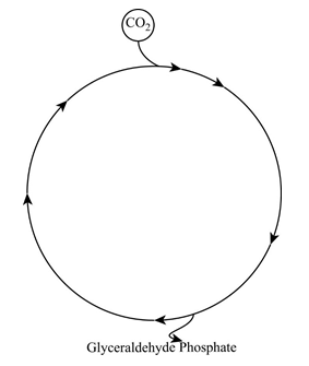
- The given diagram is the Calvin-Benson cycle. In this cycle, three molecules of carbon dioxide are fixed and one molecule of glyceraldehyde 3-phosphate is produced. After that, it leaves the cycle is shown in the below diagram.
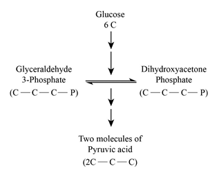
- The given b diagram resembles the Glycolysis pathway. In this pathway, the oxidation of glucose yields two molecules of pyruvic acid as its end product.
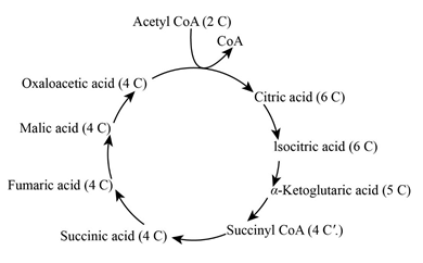
- The below-given diagram is the Kreb’s cycle. Here, the decarboxylation of pyruvic acid produces one carbon dioxide molecule and one acetyl group.
To review:
The anabolic and catabolic mechanisms in the given pathways.
Introduction:
The cellular mechanism of all living organisms requires the energy for its
Explanation of Solution
Glycolysis and the Citric acid cycle are the major pathways for anabolic and catabolic mechanisms of all organic molecules, such as carbohydrates, lipids, and proteins. Glycerol is catabolized as Dihydroxyacetone phosphate in the Glycolysis pathway (a) and fatty acids are catabolized as acetyl CoA int he TCA cycle (b).
Glutamic acid is catabolized in the Krebs cycle (c). Glutamic acid is an amino acid which is catabolized by Kreb’s cycle at α-ketoglutaric acid which is formed from isocitric acid.
In the Calvin-Benson cycle, Glyceraldehyde 3-phosphate is the end product, which enters into glycolysis. In glycolysis, the glucose is oxidized into pyruvate which is decarboxylated to acetyl group and entered into the Kreb’s cycle.
The Calvin cycle requires 18 molecules of ATP between glucose and glyceraldehyde 3- phosphate.
Three molecules of carbon dioxide (CO2) is released in the Kreb’s cycle,
- Between pyruvate to acetyl CoA.
- Between isocitric acid to α-ketoglutarate (TCA cycle).
- Between α-ketoglutarate to succinyl CoA.
The long chain hydrocarbon like such as acetyl group is catabolized in the TCA cycle at acetyl CoA. This acetyl group like hydrocarbons are catabolized by beta-oxidation which enters into the Kreb’s cycle.
The production of NADH, FADH2 or NADH in glycolysis, Calvin-Benson and TCA cycles are,
The anabolic and catabolic pathways are integrated between,
- In glycolysis, the anabolic and catabolic pathways integrated into dihydroxyacetone phosphate.
- In the TCA cycle, the anabolic and catabolic pathways integrated into acetyl, oxaloacetic acid and ketoglutaric acid.
| Utilizes | Produces | |
| Glycolysis | 2 NADH | |
| Calvin-Benson cycle | 6 NADPH | |
| Pyruvate to acetyl CoA | 1 NADH | |
| Isocitrate to α-ketoglutaric acid | 1 NADH | |
| α-keto-glutaric to succinyl CoA | 1 NADH | |
| Succinate to fumarate | 1 FADH2 | |
| Malate to oxalate | 1 NADH |
Want to see more full solutions like this?
Chapter 5 Solutions
Microbiology: An Introduction (13th Edition)


- Pleiotropic genes are genes that (blank) Cause a swapping of organs/structures, are the result of duplicated sets of chromosomes, never produce protein products, and have more than one purpose/functionarrow_forwardA loss of function mutation in Pitx1 enhancers can cause (blank) Removal of Pitx1 exons and growth of ectopic hindlimbs, growth of extra ectopic forelimbs, loss of forelimb specification and development, and loss of hindlimb specification and developmentarrow_forwardHox1a most likely contributes to (blank) patterning in the developing embryo? Ventral, posterior, limb or anteriorarrow_forward
- Select all of the following that can help establish Hox gene expression boundaries (things that affect Hox and not things that Hox affects). Retinoic acid, anterior/posterior axis, fibroblast growth factors, vagal neural crest, and enhancersarrow_forwardEctopic expression of Hox often results in (blank) phenotypes. (Blank) transformations are characterized by the replacement of one body part/structure with another. Hoxeotic, homealoneotic, joexotic, or homeoticarrow_forwardWhat's the difference when drawing omega-6 and omega-3?arrow_forward
- . Consider a base substitution mutation that occurred in a DNA sequence that resulted in a change in the encoded protein from the amino acid glutamic acid to aspartic acid. Normally the glutamic acid amino acid is located on the outside of the soluble protein but not near an active site. O-H¨ A. What type of mutation occurred? O-H B. What 2 types of chemical bonds are found in the R-groups of each amino acid? The R groups are shaded. CH2 CH2 CH2 H2N-C-COOH H2N-C-COOH 1 H Glutamic acid H Aspartic acid C. What 2 types of bonds could each R-group of each of these amino acids form with other molecules? D. Consider the chemical properties of the two amino acids and the location of the amino acid in the protein. Explain what effect this mutation will have on this protein's function and why.arrow_forwardengineered constructs that consist of hollow fibers are acting as synthetic capillaries, around which cells have been loaded. The cellular space around a single fiber can be modeled as if it were a Krogh tissue cylinder. Each fiber has an outside “capillary” radius of 100 µm and the “tissue” radius can be taken as 200 µm. The following values apply to the device:R0 = 20 µM/secaO2 = 1.35 µM/mmHgDO2,T = 1.67 x 10⁻⁵ cm2/secPO2,m = 4 x 10⁻³ cm/secInstead of blood inside the fibers, the oxygen transport and tissue consumption are being investigated by usingan aqueous solution saturated with pure oxygen. As a result, there is no mass transfer resistance in the synthetic“capillary”, only that due to the membrane itself. Rather than accounting for pO2 variations along the length ofthe fiber, use an average value in the “capillary” of 130 mmHg.Is the tissue fully oxygenated?arrow_forwardMolecular Biology Please help with question. thank you You are studying the expression of the lac operon. You have isolated mutants as described below. In the presence of glucose, explain/describe what would happen, for each mutant, to the expression of the lac operon when you add lactose AND what would happen when the bacteria has used up all of the lactose (if the mutant is able to use lactose).5. Mutations in the lac operator that strengthen the binding of the lac repressor 200 fold 6. Mutations in the promoter that prevent binding of RNA polymerase 7. Mutations in CRP/CAP protein that prevent binding of cAMP8. Mutations in sigma factor that prevent binding of sigma to core RNA polymerasearrow_forward
- Molecular Biology Please help and there is an attached image. Thank you. A bacteria has a gene whose protein/enzyme product is involved with the synthesis of a lipid necessary for the synthesis of the cell membrane. Expression of this gene requires the binding of a protein (called ACT) to a control sequence (called INC) next to the promoter. A. Is the expression/regulation of this gene an example of induction or repression?Please explain:B. Is this expression/regulation an example of positive or negative control?C. When the lipid is supplied in the media, the expression of the enzyme is turned off.Describe one likely mechanism for how this “turn off” is accomplished.arrow_forwardMolecular Biology Please help. Thank you. Discuss/define the following:(a) poly A polymerase (b) trans-splicing (c) operonarrow_forwardMolecular Biology Please help with question. Thank you in advance. Discuss, compare and contrast the structure of promoters inprokaryotes and eukaryotes.arrow_forward

 Human Heredity: Principles and Issues (MindTap Co...BiologyISBN:9781305251052Author:Michael CummingsPublisher:Cengage Learning
 Concepts of BiologyBiologyISBN:9781938168116Author:Samantha Fowler, Rebecca Roush, James WisePublisher:OpenStax College