
Videos
Connecting the Concepts
1. Use the following diagram to review the flow of blood through a human cardiovascular system. Label the indicated parts, highlight the vessels that carry oxygen-rich blood, and then trace the flow of blood by numbering the circles from 1 to 10, starting with 1 in the right ventricle. (When two locations are equivalent in the pathway, such as right and left lung capillaries or capillaries of top and lower portion of the body, assign them the same number.)
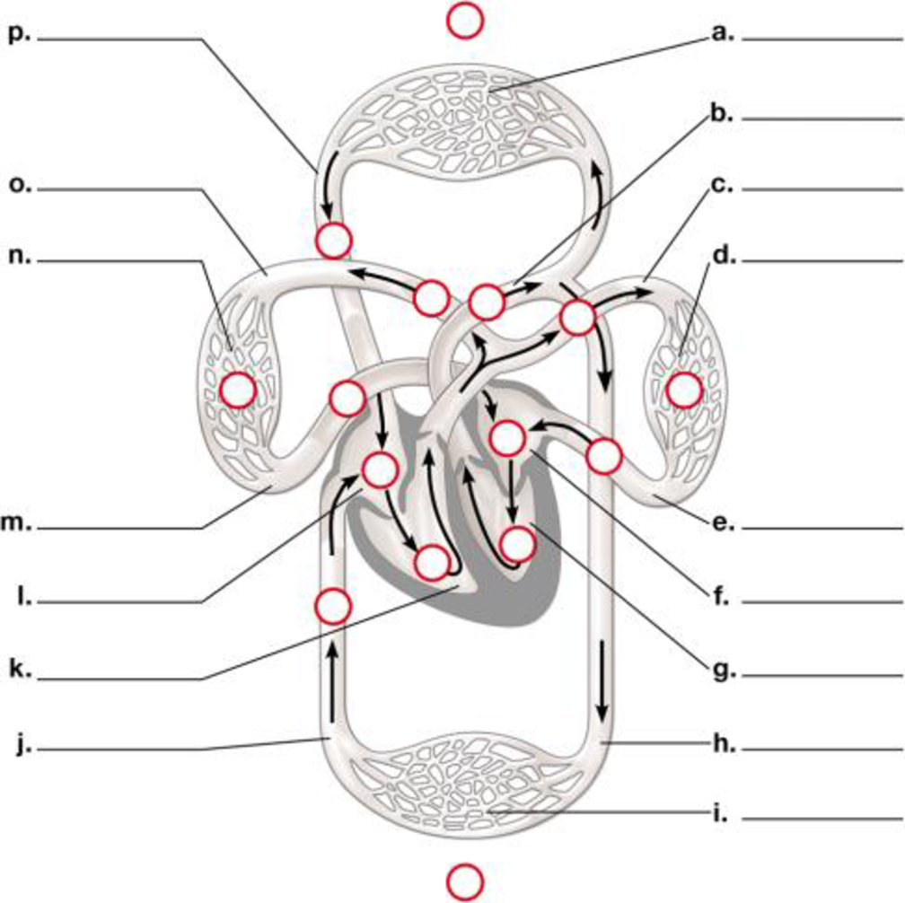
To label: The diagram of the flow of blood through the circulatory system.
Concept introduction:
The blood flows through the circulatorysystem. It includes various capillaries, aorta, pulmonary artery, pulmonary veins, ventricles, auricles, superior and inferior vena cava.
Answer to Problem 1CC
Pictorial representation: A labelled diagram of human circulatory system showing the flow of blood through it is given on Fig.1.

Fig. 1: The circulatory system.
Explanation of Solution
The labeling of the blood vessels in the circulatory system include as follows:
(a)
Network of the blood capillaries: It supplies blood to the various parts of the human body like head, arms, chest and legs.
(b)
Aorta: The blood moves from the heart chambers through this blood vessel.
(c)
Pulmonary artery: It carries the deoxygenated blood.
(d)
Capillaries: They supply blood to the lungs: The blood flows through them and reaches the lungs.
(e)
Pulmonary vein: It carries oxygenated blood.
(f)
Left atrium: It is the upper chamber of the heart which contracts to cause the flow of the blood out of the heart.
(g)
Left ventricle: It is the lower chamber of the heart where the blood flows.
(h)
Aorta: This blood vessel transports the blood to the organs from the heart.
(i)
Capillaries: They supply the blood of abdominal regions and legs:
(j)
Inferior vena cava: It drains the blood into the right atrium.
(k)
Right ventricle: It is the lower chamber of the heart.
(l)
Right atrium: It is the upper chamber of the heart.
(m)
Pulmonary vein: The left atrium of the heart receives blood from the pulmonary vein.
(n)
Capillaries of right lung: It helps in the flow of the blood to the lungs.
(o)
Pulmonary artery: It carries deoxygenated blood.
(p)
Superior vena cava: It circulates the blood in the sino-atrial node in the right atrium.
Want to see more full solutions like this?
Chapter 23 Solutions
Campbell Biology: Concepts & Connections (9th Edition)


- 22. Which of the following mutant proteins is expected to have a dominant negative effect when over- expressed in normal cells? a. mutant PI3-kinase that lacks the SH2 domain but retains the kinase function b. mutant Grb2 protein that cannot bind to RTK c. mutant RTK that lacks the extracellular domain d. mutant PDK that has the PH domain but lost the kinase function e. all of the abovearrow_forwardWhat is the label ?arrow_forwardCan you described the image? Can you explain the question as well their answer and how to get to an answer to an problem like this?arrow_forward
- Describe the principle of homeostasis.arrow_forwardExplain how the hormones of the glands listed below travel around the body to target organs and tissues : Pituitary gland Hypothalamus Thyroid Parathyroid Adrenal Pineal Pancreas(islets of langerhans) Gonads (testes and ovaries) Placentaarrow_forwardWhat are the functions of the hormones produced in the glands listed below: Pituitary gland Hypothalamus Thyroid Parathyroid Adrenal Pineal Pancreas(islets of langerhans) Gonads (testes and ovaries) Placentaarrow_forward
 Human Physiology: From Cells to Systems (MindTap ...BiologyISBN:9781285866932Author:Lauralee SherwoodPublisher:Cengage Learning
 Biology 2eBiologyISBN:9781947172517Author:Matthew Douglas, Jung Choi, Mary Ann ClarkPublisher:OpenStax
 Anatomy & PhysiologyBiologyISBN:9781938168130Author:Kelly A. Young, James A. Wise, Peter DeSaix, Dean H. Kruse, Brandon Poe, Eddie Johnson, Jody E. Johnson, Oksana Korol, J. Gordon Betts, Mark WomblePublisher:OpenStax College
 Fundamentals of Sectional Anatomy: An Imaging App...BiologyISBN:9781133960867Author:Denise L. LazoPublisher:Cengage Learning