
Videos
Connecting the Concepts
1. Use the following diagram to review the flow of blood through a human cardiovascular system. Label the indicated parts, highlight the vessels that carry oxygen-rich blood, and then trace the flow of blood by numbering the circles from 1 to 10, starting with 1 in the right ventricle. (When two locations are equivalent in the pathway, such as right and left lung capillaries or capillaries of top and lower portion of the body, assign them the same number.)
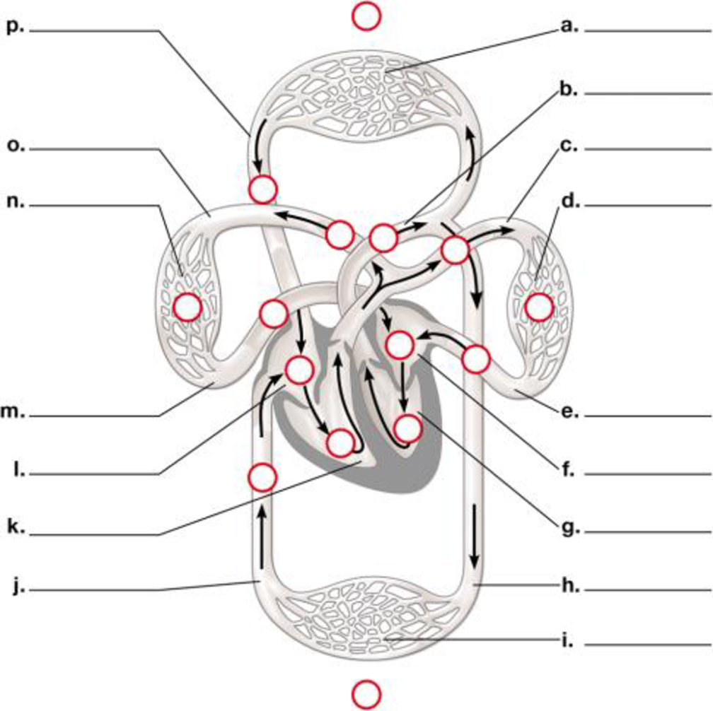
To label: The diagram of the flow of blood through the circulatory system.
Concept introduction:
The blood flows through the circulatorysystem. It includes various capillaries, aorta, pulmonary artery, pulmonary veins, ventricles, auricles, superior and inferior vena cava.
Answer to Problem 1CC
Pictorial representation: A labelled diagram of human circulatory system showing the flow of blood through it is given on Fig.1.

Fig. 1: The circulatory system.
Explanation of Solution
The labeling of the blood vessels in the circulatory system include as follows:
(a)
Network of the blood capillaries: It supplies blood to the various parts of the human body like head, arms, chest and legs.
(b)
Aorta: The blood moves from the heart chambers through this blood vessel.
(c)
Pulmonary artery: It carries the deoxygenated blood.
(d)
Capillaries: They supply blood to the lungs: The blood flows through them and reaches the lungs.
(e)
Pulmonary vein: It carries oxygenated blood.
(f)
Left atrium: It is the upper chamber of the heart which contracts to cause the flow of the blood out of the heart.
(g)
Left ventricle: It is the lower chamber of the heart where the blood flows.
(h)
Aorta: This blood vessel transports the blood to the organs from the heart.
(i)
Capillaries: They supply the blood of abdominal regions and legs:
(j)
Inferior vena cava: It drains the blood into the right atrium.
(k)
Right ventricle: It is the lower chamber of the heart.
(l)
Right atrium: It is the upper chamber of the heart.
(m)
Pulmonary vein: The left atrium of the heart receives blood from the pulmonary vein.
(n)
Capillaries of right lung: It helps in the flow of the blood to the lungs.
(o)
Pulmonary artery: It carries deoxygenated blood.
(p)
Superior vena cava: It circulates the blood in the sino-atrial node in the right atrium.
Want to see more full solutions like this?
Chapter 23 Solutions
CAMPBELL BIO: CONCEPTS&CONNECTIONS (LL)


- 9 S es Read the section "Investigating Life: In (Extremely) Cold Blood." Then, drag and drop the terms on the left to complete the concept map. Red blood cells Genes Icefishes -have mutated have colorless Oxygen have few lack encode Blood Cellular respiration consists of- contain carries is a Platelets White blood cells carries low amounts of Hemoglobin is necessary for Plasma Protein Reset.arrow_forwardPlating 50 microliters of a sample diluted by a factor of 10-6 produced 91 colonies. What was the originalcell density (CFU/ml) in the sample?arrow_forwardEvery tutor here has got this wrong, don't copy off them.arrow_forward
- Suppose that the population from question #1 (data is in table below) is experiencing inbreeding depression (F=.25) (and no longer experiencing natural selection). Calculate the new expected genotype frequencies (f) in this population after one round of inbreeding. Please round to 3 decimal places. Genotype Adh Adh Number of Flies 595 Adh Adh 310 Adhs Adhs 95 Total 1000 fladh Adh- flAdn Adh fAdhs Adharrow_forwardWhich of the following best describes why it is difficult to develop antiviral drugs? Explain why. A. antiviral drugs are very difficult to develop andhave no side effects B. viruses are difficult to target because they usethe host cell’s enzymes and ribosomes tometabolize and replicate C. viruses are too small to be targeted by drugs D. viral infections usually clear up on their ownwith no problemsarrow_forwardThis question has 3 parts (A, B, & C), and is under the subject of Nutrition. Thank you!arrow_forward
- They got this question wrong the 2 previous times I uploaded it here, please make sure it's correvct this time.arrow_forwardThis question has multiple parts (A, B & C), and under the subject of Nutrition. Thank you!arrow_forwardCalculate the CFU/ml of a urine sample if 138 E. coli colonies were counted on a Nutrient Agar Plate when0.5 mls were plated on the NA plate from a 10-9 dilution tube. You must highlight and express your answerin scientific notatioarrow_forward
 Human Physiology: From Cells to Systems (MindTap ...BiologyISBN:9781285866932Author:Lauralee SherwoodPublisher:Cengage Learning
 Biology 2eBiologyISBN:9781947172517Author:Matthew Douglas, Jung Choi, Mary Ann ClarkPublisher:OpenStax
 Anatomy & PhysiologyBiologyISBN:9781938168130Author:Kelly A. Young, James A. Wise, Peter DeSaix, Dean H. Kruse, Brandon Poe, Eddie Johnson, Jody E. Johnson, Oksana Korol, J. Gordon Betts, Mark WomblePublisher:OpenStax College
 Fundamentals of Sectional Anatomy: An Imaging App...BiologyISBN:9781133960867Author:Denise L. LazoPublisher:Cengage Learning