
Concept explainers
Videos
To write:
About some diseases caused by protozoans and their location on map.
Introduction:
Protozoans are animal-like protists who follow a heterotrophic mode of nutrition and are thus characterized as heterotrophs. In the heterotrophic mode of nutrition, an organism is unable to synthesize their food and depend on others for obtaining nutrients and energy. Protozoans generally ingest other protozoans, algae, and bacteria to obtain energy and nutrition.
Explanation of Solution
Some of the common diseases caused by protozoans include malaria, African sleeping sickness, amoebic dysentery, and leishmaniasis.
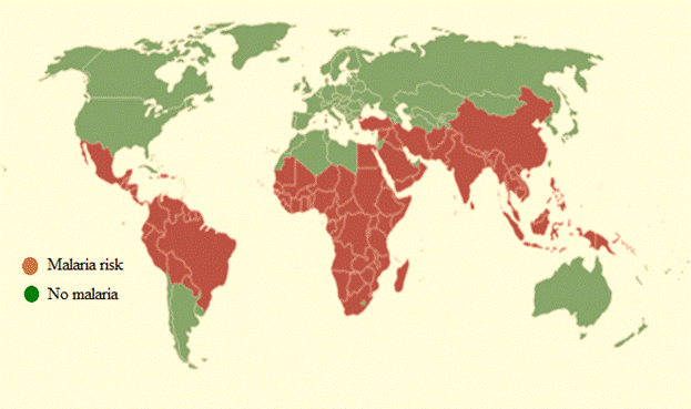
Malaria is caused by Plasmodium which is transmitted by the bite of a female Anopheles mosquito which acts as an intermediate host of plasmodium. Malaria is generally spread in subtropical and tropical regions where conditions like rainfall, humidity, and high temperature favor the plasmodium and mosquito growth. The breeding ground of Anopheles mosquito and plasmodium includes stagnant water which can be found in tyre tracks, hoof prints, pond margins, seepages, rice fields, irrigation channels, rainwater pools, burrow pits, and wells. The map showing locations of malaria affected regions is given below:
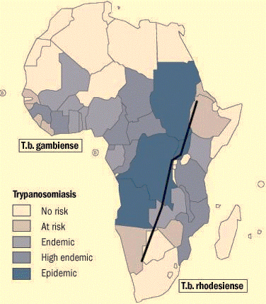
African sleeping sickness is caused by Trypanosoma brucei and is transmitted by tsetse fly. This disease is specifically diagnosed in regions of sub-Saharan Africa. Symptoms of sleeping sickness include itching, joint pains, headache, and fever. The map showing locations of sleeping sickness affected regions is given below:
Amoebic dysentery is caused by amoeba and is spread through intake of contaminated food and water. Abdomen is majorly affected in this infection and other symptoms include flatulence, diarrhoea, and blood in stool. This infection is generally spotted in regions of Asia, Africa, South America, and Central America.The map showing locations of amoebic dysentery affected regions is given below:
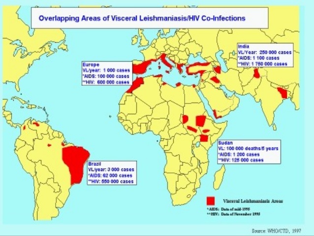
Leishmaniasis is caused by Leishmania type
Chapter 19 Solutions
Glencoe Biology, Florida Edition
Additional Science Textbook Solutions
Physics for Scientists and Engineers: A Strategic Approach, Vol. 1 (Chs 1-21) (4th Edition)
Applications and Investigations in Earth Science (9th Edition)
Cosmic Perspective Fundamentals
Anatomy & Physiology (6th Edition)
Campbell Biology (11th Edition)
Organic Chemistry (8th Edition)


- Please finish the chart at the bottom. Some of the answers have been filled in.arrow_forward9. Aerobic respiration of one lipid molecule. The lipid is composed of one glycerol molecule connected to two fatty acid tails. One fatty acid is 12 carbons long and the other fatty acid is 18 carbons long in the figure below. Use the information below to determine how much ATP will be produced from the glycerol part of the lipid. Then, in part B, determine how much ATP is produced from the 2 fatty acids of the lipid. Finally put the NADH and ATP yields together from the glycerol and fatty acids (part A and B) to determine your total number of ATP produced per lipid. Assume no other carbon source is available. 18 carbons fatty acids 12 carbons 9 glycerol A. Glycerol is broken down to glyceraldehyde 3-phosphate, a glycolysis intermediate via the following pathway shown in the figure below. Notice this process costs one ATP but generates one FADH2. Continue generating ATP with glyceraldehyde-3-phosphate using the standard pathway and aerobic respiration. glycerol glycerol-3- phosphate…arrow_forwardNormal dive (for diving humans) normal breathing dive normal breathing Oz level CO2 level urgent need to breathe Oz blackout zone high CO2 triggers breathing 6. This diagram shows rates of oxygen depletion and carbon dioxide accumulation in the blood in relation to the levels needed to maintain consciousness and trigger the urgent need to breathe in diving humans. How might the location and slope of the O₂ line differ for diving marine mammals such as whales and dolphins? • How might the location and slope of the CO₂ line differ for diving marine mammals such as whales and dolphins? • • Draw in predicted lines for O2 and CO2, based on your reasoning above. How might the location of the Urgent Need to Breathe line and the O2 Blackout Zone line differ for diving marine mammals? What physiological mechanisms account for each of these differences, resulting in the ability of marine mammals to stay submerged for long periods of time?arrow_forward
- foraging/diet type teeth tongue stomach intestines cecum Insectivory numerous, spiky, incisors procumbentExample: moleExample: shrew -- simple short mostly lacking Myrmecophagy absent or reduced in numbers, peg-likeExample: tamandua anteater extremely long simple, often roughened short small or lacking Terrestrial carnivory sharp incisors; long, conical canines; often carnassial cheek teeth; may have crushing molarsExample: dog -- simple short small Aquatic carnivory homodont, spiky, numerousExample: common dolphin -- simple or multichambered (cetaceans only) variable small or absent Sanguinivory very sharp upper incisors; reduced cheek teethExample: vampire bat grooved tubular, highly extensible long small or lacking Herbivory (except nectivores) incisors robust or absent; canines reduced or absent; diastema; cheek teeth enlarged with complex occlusal surfacesExample: beaver -- simple (hindgut fermenters) or multichambered (ruminants) long large Filter feeding none…arrow_forward3. Shown below is the dental formula and digestive tract anatomy of three mammalian species (A, B, and C). What kind of diet would you expect each species to have? Support your answers with what you can infer from the dental formula and what you can see in the diagram. Broadly speaking, what accounts for the differences? Species A 3/3, 1/1, 4/4, 3/3 པར『ན་ cm 30 Species B 4/3, 1/1, 2/2, 4/4 cm 10 Species C 0/4, 0/0,3/3, 3/3 020arrow_forward3. Shown below is the dental formula and digestive tract anatomy of three mammalian species (A, B, and C). What kind of diet would you expect each species to have? Support your answers with what you can infer from the dental formula and what you can see in the diagram. Broadly speaking, what accounts for the differences? Species A 3/3, 1/1, 4/4, 3/3 cm 30 Species B 0/4, 0/0, 3/3, 3/3 cm 10 Species C 4/3, 1/1, 2/2, 4/4 E 0 cm 20 AILarrow_forward
- Normal dive (for diving humans) normal breathing dive normal breathing Oz level CO₂ level urgent need to breathe Oz blackout zone high CO₂ triggers breathing 6. This diagram shows rates of oxygen depletion and carbon dioxide accumulation in the blood in relation to the levels needed to maintain consciousness and trigger the urgent need to breathe in diving humans. • How might the location and slope of the O2 line differ for diving marine mammals such as whales and dolphins? • How might the location and slope of the CO2 line differ for diving marine mammals such as whales and dolphins? • • Draw in predicted lines for O2 and CO2, based on your reasoning above. How might the location of the Urgent Need to Breathe line and the O2 Blackout Zone line differ for diving marine mammals? What physiological mechanisms account for each of these differences, resulting in the ability of marine mammals to stay submerged for long periods of time?arrow_forwardHow much ATP will be produced during the following metabolic scenario: Aerobic respiration of a 5mM lipid solution that is made up of one glycerol and an 8-carbon fatty acid and 12-carbon fatty acid. Recall that when glycerol breaks down to Glyceraldehyde-3-phosphate it costs one ATP but your get an extra FADH2. Every two carbons of a fatty acid break down to one acetyl-CoA. Units cannot be entered in this style of question but the units of your answer should be in mM of ATP.arrow_forwardIf a bacterium using aerobic respiration was to degrade one small protein molecule into 8 molecules of pyruvic acid, how many ATP would that cell make? Assume there is no other carbon source. Units cannot be entered in this style of question but the units of your answer should be in molecules of ATP.arrow_forward
- If a bacterium using aerobic respiration was to degrade a 30 mM solution of citric acid, how many ATP would that cell make? Assume no other carbon source is available. Units cannot be entered in this style of question but the units of your answer should be in mM of ATP.arrow_forwardHow much ATP will be produced during the following metabolic scenario: Aerobic respiration of a 5mM lipid solution that is made up of one glycerol and an 8-carbon fatty acid and 12-carbon fatty acid. Recall that when glycerol breaks down to Glyceraldehyde-3-phosphate it costs one ATP but your get an extra FADH2. Every two carbons of a fatty acid break down to one acetyl-CoA. (pathways will be provided on the exam) Units cannot be entered in this style of question but the units of your answer should be in mM of ATP.arrow_forwardWhen beta-lactamase was isolated from Staphylcoccus aureus and treated with a phosphorylating agent, only the active site, serine was phosphorylated. Additionally, the serine was found to constitute 0.35% (by weight) of this beta-lactamase enzyme. Using this, calculate the molecular weight of this enzyme and estimate the number of amino acids present in the polypeptide.arrow_forward
 Human Anatomy & Physiology (11th Edition)BiologyISBN:9780134580999Author:Elaine N. Marieb, Katja N. HoehnPublisher:PEARSON
 Biology 2eBiologyISBN:9781947172517Author:Matthew Douglas, Jung Choi, Mary Ann ClarkPublisher:OpenStax
 Anatomy & PhysiologyBiologyISBN:9781259398629Author:McKinley, Michael P., O'loughlin, Valerie Dean, Bidle, Theresa StouterPublisher:Mcgraw Hill Education,
 Molecular Biology of the Cell (Sixth Edition)BiologyISBN:9780815344322Author:Bruce Alberts, Alexander D. Johnson, Julian Lewis, David Morgan, Martin Raff, Keith Roberts, Peter WalterPublisher:W. W. Norton & Company
 Laboratory Manual For Human Anatomy & PhysiologyBiologyISBN:9781260159363Author:Martin, Terry R., Prentice-craver, CynthiaPublisher:McGraw-Hill Publishing Co.
 Inquiry Into Life (16th Edition)BiologyISBN:9781260231700Author:Sylvia S. Mader, Michael WindelspechtPublisher:McGraw Hill Education