To label: The structures given in Fig 18.1
Concept introduction: Neurons are the fundamental functioning units of the nervous system. Neurons share many similar features with other body cells; only one different feature is that they can conduct signals throughout the body.
Answer to Problem 1.1BGL
Pictorial representation:

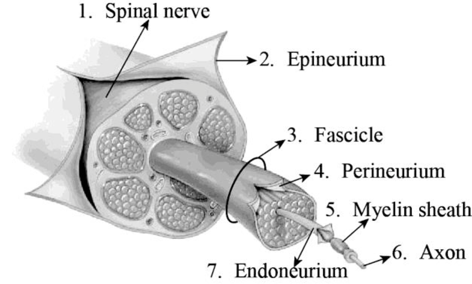
Fig 1: Transverse section displaying the coverings of a spinal nerve
Explanation of Solution
The spinal nerves function like “telephone lines” and bring back and forth messages between the body and the spinal cord to regulate motions as well as sensations. There are two roots in each spinal nerve; they are the ventral root and the dorsal root. An axon is a lengthy, slender projection that emerges from a nerve cell. The myelin sheath is formed of a fatty material that surrounds the nerve cells.
The dense and uneven connective tissue that surrounds the peripheral nerves is termed as the epineurium. This tissue envelops several blood vessels that supply blood to the nerves and nerve fascicles. The epineurium is abundantly seen in the regions of joints. Hence, they protect the nerves from injury as well as stretching.
A nerve fascicle is otherwise known as fasciculus. This structure is formed of a bundle of axons. These fascicles are present in the peripheral nervous system. They are bundled together with the blood vessels and fatty tissues.
Nerve fascicles are enveloped by a kind of connective tissue that is termed as the perineurium. The perineurium is formed of perineurial cells, and they are also referred to as myoepithelioid in some cases. The perineurium membrane is a smooth, tube-like transparent structure that could be easily detached from the fibers which it surrounds.
The connective tissue that surrounds the unmyelinated and myelinated axons is called the endoneurium. The endoneurium is otherwise known as the endoneurial channel or Henle’s sheath. It is delicate and surrounds the myelin sheath of every myelinated nerve fiber. The endoneurium is made up of endoneurial cells.
To label: The structures given in Fig 18.2
Answer to Problem 1.1BGL
Pictorial representation:

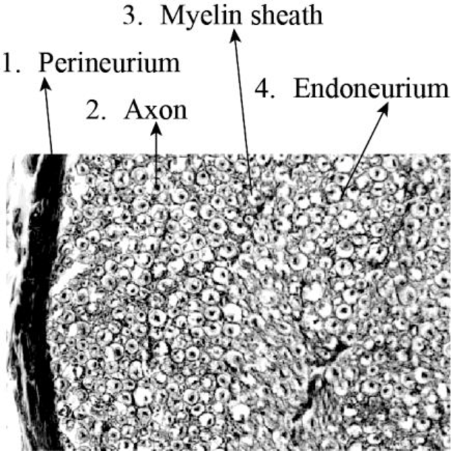
Fig 2: A transverse section photomicrograph through a spinal nerve fascicle
Explanation of Solution
An axon is a long, slim projection that emerges from a neuron. The myelin sac consists of a fat material that surrounds the cells of the nerves. The endoneurium is also known as the Henle’s sheath or endoneurial channel. It is fragile and surrounds every myelinated nerve fiber’s sheath. The endoneurium consists of endoneurial cells.
Want to see more full solutions like this?
Chapter 18 Solutions
Laboratory Manual for Anatomy and Physiology, 6e WileyPLUS (next generation) + Loose-leaf
- View History Bookmarks Window Help Quarter cements ents ons (17) YouTube Which amino acids would you expect to find marked on the alpha helix? canvas.ucsc.edu ucsc Complaint and Grievance Process - Academic Personnel pach orations | | | | | | | | | | | | | | | | 000000 000000000 | | | | | | | | | | | | | | | | | | | | | | 00000000 scope vious De 48 12.415 KATPM FEB 3 F1 F2 80 F3 a F4 F5 2 # 3 $ 85 % tv N A の Mon Feb 3 10:24 PM Lipid bilayer Submit Assignment Next > ZOOM < Å DII 8 བ བ F6 16 F7 F8 F9 F10 34 F11 F12 & * ( 6 7 8 9 0 + 11 WERTY U { 0 } P deletearrow_forwardDifferent species or organisms research for ecologyarrow_forwardWhat is the result of the following gram stain: positive ○ capsulated ○ acid-fast ○ negativearrow_forward
- What type of stain is the image below: capsule stain endospore stain gram stain negative stain ASM MicrobeLibrary.org Keplingerarrow_forwardWhat is the result of the acid-fast stain below: Stock Images by Getty Images by Getty Images by Getty Images by Getty Image Getty Images St Soy Getty Images by Getty Images by Getty Images Joy Getty encapsulated O endosporulating negative ○ positivearrow_forwardYou have a stock vial of diligence 75mg in 3ml and need to draw up a dose of 50mg for your patient.how many mls should you draw up to give this dosearrow_forward
- You are recquired to administer 150mg hydrocortisone intravenously,how many mls should you give?(stock =hydrocortisone 100mg in 2mls)arrow_forwardIf someone was working with a 50 MBq F-18 source, what would be the internal and external dose consequences?arrow_forwardWe will be starting a group project next week where you and your group will research and ultimately present on a current research article related to the biology of a pathogen that infects humans. The article could be about the pathogen itself, the disease process related to the pathogen, the immune response to the pathogen, vaccines or treatments that affect the pathogen, or other biology-related study about the pathogen. I recommend that you choose a pathogen that is currently interesting to researchers, so that you will be able to find plenty of articles about it. Avoid choosing a historical disease that no longer circulates. List 3 possible pathogens or diseases that you might want to do for your group project.arrow_forward
- not use ai pleasearrow_forwardDNK dagi nukleotidlar va undan sintezlangan oqsildagi peptid boglar farqi 901 taga teng bo'lib undagi A jami H boglardan 6,5 marta kam bo'lsa DNK dagi jami H bog‘lar sonini topingarrow_forwardOne of the ways for a cell to generate ATP is through the oxidative phosphorylation. In oxidative phosphorylation 3 ATP are produced from every one NADH molecule. In respiration, every glucose molecule produces 10 NADH molecules. If a cell is growing on 5 glucose molecules, how much ATP can be produced using oxidative phosphorylation/aerobic respiration?arrow_forward
 Human Anatomy & Physiology (11th Edition)BiologyISBN:9780134580999Author:Elaine N. Marieb, Katja N. HoehnPublisher:PEARSON
 Biology 2eBiologyISBN:9781947172517Author:Matthew Douglas, Jung Choi, Mary Ann ClarkPublisher:OpenStax
 Anatomy & PhysiologyBiologyISBN:9781259398629Author:McKinley, Michael P., O'loughlin, Valerie Dean, Bidle, Theresa StouterPublisher:Mcgraw Hill Education,
 Molecular Biology of the Cell (Sixth Edition)BiologyISBN:9780815344322Author:Bruce Alberts, Alexander D. Johnson, Julian Lewis, David Morgan, Martin Raff, Keith Roberts, Peter WalterPublisher:W. W. Norton & Company
 Laboratory Manual For Human Anatomy & PhysiologyBiologyISBN:9781260159363Author:Martin, Terry R., Prentice-craver, CynthiaPublisher:McGraw-Hill Publishing Co.
 Inquiry Into Life (16th Edition)BiologyISBN:9781260231700Author:Sylvia S. Mader, Michael WindelspechtPublisher:McGraw Hill Education


