To match: The terms with the organs/structures of the
Introduction: The digestive system consists of a collection of organs that help in the digestion of food materials and converting them into basic energy rich molecules for supplying it to the entire body.
Answer to Problem 1P
Pictorial representation: The organs or structures of the digestive system are given in Fig. 1.
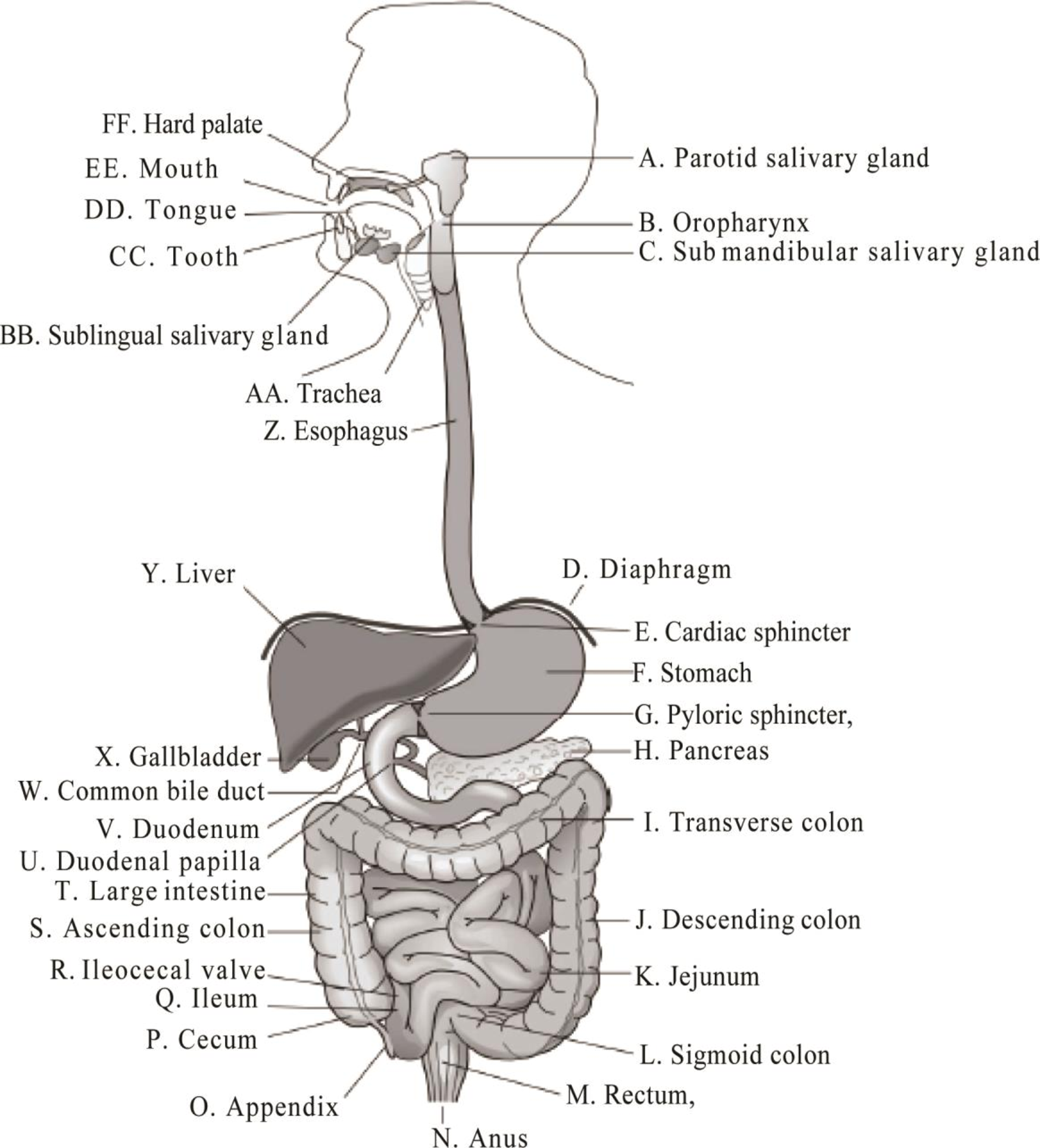
Fig.1: The organs or structures of the digestive system
Explanation of Solution
The human digestive system is formed of gastrointestinal tract and accessory organs of digestion.
A. Parotid salivary gland: The parotid salivary gland secretes the serous saliva into the mouth through the parotid duct, to facilitate mastication and swallowing and to begin the digestion of starches.
B. Oropharynx: The muscular walls of it functions in the process of swallowing, and also it serves as a pathway for the movement of food from the mouth to the esophagus.
C. Sub-mandibular salivary gland: The sub-mandibular gland is one of the major glands that provide the mouth with saliva containing digestive enzymes that moistens the food.
D. Diaphragm: The dome-shaped muscle that divides the thoracic and abdominal cavities and involved in the respiration process is known as diaphragm. The diaphragm becomes expand when the lungs are in the relaxed state during excretion. It becomes flatten when the lungs expand for inhalation.
E. Cardiac sphincter: The cardiac sphincter or lower sphincter is located at the upper portion of the stomach. It prevents the acidic contents of the stomach from moving upward into the esophagus.
F. Stomach: The stomach is a muscular sac that is expansible and acts as the food and fluid reservoir. The food from the esophagus is received by the stomach. Gastric acids and digestive enzymes for the digestion of food are secreted in the stomach.
G. Pyloric sphincter: The pyloric sphincter forms a junction between the pylorus of the stomach and the duodenum of the small intestine. It acts as a valve to control the flow of partially digested food from the stomach to the small intestine.
H. Pancreas: The pancreas has a loose, lumpy structure and is made up of glandular tissues. It secretes the digestive enzymes into the small intestine. The islets of Langerhans are the region present in the pancreas, which consists of endocrine cells. The islets of Langerhans will produce and secrete hormones.
I. Transverse colon: The transverse colon connects the ascending colon to the descending colon. It moves the waste material to the rectum by peristalsis and haustral churning. It absorbs water and electrolytes from the digested food.
J. Descending colon: The descending colon is the part of the large intestine. The function of descending colon is to store the remains of digested food that will be transferred into the rectum.
K. Jejunum: The jejunum is the part of the intestine. The main function of jejunum is to absorb nutrients from the digested food.
L. Sigmoid colon: The sigmoid colon is a part of the large intestine that connects the descending colon to the rectum. Its main function is to store fecal wastes until defecation.
M. Rectum: The rectum is the chamber that begins at the end of the large intestine; it receives the stool from the descending colon and produces an urge to defecate.
N. Anus: The anus is the last part of the digestive tract. Its function is to control the expulsion of feces and the unwanted semi-solid matter produced during digestion.
O. Appendix: The appendix (vermiform appendix) is situated in the lower right side of the abdomen. The function of the appendix is unknown.
P. Cecum: The main function of the cecum is to absorb salts and fluids that remain after the completion of intestinal digestion and absorption and to mix it with mucus.
Q. Ileum: The ileum is the part of the intestine. It absorbs vitamin B12, bile salts, and the other products, which are not absorbed by the jejunum.
R. Ileocecal valve: The ileocecal valve is the joining point between the large and small intestine. It serves as a barrier to prevent bacteria laden contents from large intestine to the small intestine.
S. Ascending colon: The ascending colon is a part of the large intestine. It carries feces from the cecum to the transverse colon.
T. Large intestine: The large intestine is the last part of the gastrointestinal tract. The main functions of the large intestine are the recovery of water and electrolytes, formation and storage of feces, fermentation of indigestible food by bacteria.
U. Duodenal papilla: The secretions from the liver and the exocrine pancreas are added to the chime in the duodenum through the duodenal papilla for the chemical digestion of food.
V. Duodenum: The duodenum is the upper part of the small intestine where the chemical secretions from the pancreas, liver, and gallbladder mix with the chime to facilitate the chemical digestion.
W. Common bile duct: The common bile duct is a small structure present in the region where the common hepatic duct and the cystic duct join. Its physiological role is to carry bile from the gallbladder to the duodenum.
X. Gallbladder: The gallbladder is one of the additional organs that support the digestive process. The gallbladder helps to store bile (a thick yellow fluid produced by the liver that helps for lipid digestion) before its release into the small intestine. When the composition of bile increases from the normal level, it starts to accumulate in the gallbladder and form gallstones.
Y. Liver: Liver is the largest gland in the human body that is often referred to as “
Z. Esophagus: The esophagus forms an important part of the gastrointestinal tract. It is thin, long, and muscular tube that connects the pharynx to the stomach.
AA. Trachea: The trachea is a windpipe also called throat, the pharynx is the part of the digestive tract that receives food from the mouth and carries to the esophagus.
BB. Sublingual salivary gland: The sublingual salivary glands are present under the mucus membrane. It secretes the saliva containing digestive enzymes that moisten the food and begin to break down food before it passes into the stomach.
CC. Tooth: The tooth is the hard, calcified structure that is present in the mouth teeth. Tooth helps in the mastication process. The process by which the food is crushed and ground by teeth is called mastication process. The food will break into pieces by chewing and it will easily get digested.
DD. Tongue: The tongue is the muscular organ present inside the mouth. It is covered with mucus and thousands of taste buds are present on the surfaces of the papillae. Tongue and teeth in the mouth will start the digestion process by chewing the food.
EE. Mouth: The mouth plays vital role in drinking, eating, breathing, and speaking. Tongue, teeth, and salivary glands are present inside the mouth; the digestion process starts at the mouth by breaking the food into pieces by chewing and mixing with saliva.
FF. Hard palate: The hard palate is present as a partition between the nasal passages and the mouth. It facilitates the movement of food backward towards the larynx.
Want to see more full solutions like this?
Chapter 17 Solutions
Study Guide for Gould's Pathophysiology for the Health Professions
- Objective: Develop a culturally sensitive nursing care plan by addressing a hypothetical patient’s cultural beliefs, values, and practices that influence their health and healthcare decisions. Instructions: 1. Scenario Analysis: o Carefully read the provided hypothetical patient scenario. o Identify cultural, religious, or personal beliefs that may aJect the patient’s health behaviors and decision-making. 2. Cultural Assessment: o Conduct a cultural assessment based on the scenario. Include the patient’s cultural background, health beliefs, communication preferences, dietary restrictions, and practices related to illness and healing. 3. Nursing Care Plan Components: o Assessment: Identify the patient’s main health concerns and cultural needs. o Diagnosis: Formulate culturally sensitive nursing diagnoses. o Goals and Outcomes: Establish realistic, measurable goals that respect the patient’s cultural preferences. o Interventions: Propose specific nursing interventions that accommodate…arrow_forwardOne of your long-term patients who you have known for many years has progressed to end-stage prostate cancer and has been placed on a palliative care program. The currently commercially available morphine liquids he has been using contain a flavouring agent that makes him nauseous. His Physician has requested you compound a morphine liquid for him without flavour as his pain is well controlled on this medication and he does not want to change to another pain reliever. Your pharmacy team and the Physician would like to make his end-of-life process as comfortable as possible. A formulation for a suspension appears to be a good option to try. RX: Morphine liquid 1 mg/mL Sig: Take 1-2 mL q1h prn Mitte: 100 mL Formulation: Morphine HCl 10 mg Glycerol 1 mL Compound Hydroxybenzoate Solution 0.1 mL Purified water to 10 mL Use within 1 montharrow_forwardAs a nursing student in the pediatric unit, explore the topic "fear and anxiety during hospitalization for school age patients" in a TGROW format. Topic: What issue are you planning to address? Provide overview.Goal: What is your goal? Is it SMART?Reality: Current state of the situation? Why are you choosing this goal? What hashappened?Options: What are all the possible options to deal with the situation? 3 should bearticulated. What obstacles might be in the way? Weigh the pros and cons of eachoption.Way Forward: Which option are you selecting? What do you need to get done to achieveyour goal? What will be your first step? Commit to taking action.arrow_forward
- why is it important to understand children experiencing fear and anxiety while being admitted to the hospital as a nursing student and how does this impact nursing practicearrow_forwardAs a nursing student in the pediatric unit, explore the topic "fear and anxiety during hospitalization for school age patients" in a TGROW formatarrow_forwardwhat are essential skills of nursingarrow_forward
- As a nursing student in the pediatric unit, what is an example of a TGROW with a Topic: Fears and anxiety in pediatric patients during hospitalization. What can be the Goal (SMART), Reality, Options and Way forwardarrow_forwardAs a nursing student, provide 5 examples of a TGROW that is relevant in the pediatric unit.arrow_forwardwhat are therapies needed for mechanicaly ventilated patients?arrow_forward
- What therapies are indicated for a patient on invasive mechanical ventilation? What weaning parameters should be performed and what are acceptable values?arrow_forwardDomains of Enterprise Risks 1. Strategic- Examples of risks: mergers, acquisitions, brand/reputation 2. Operational- Examples of risks: efficiencies, following policies/procedures, equitable care 3. Financial- Examples of risks: capital, cost of malpractice claims, claims fraud/abuse, billing Describe each enterprise risk domain Describe a potential risk for each domain Analyze potential outcomes associated with each risk Recommend actions to avoid each risk (proactively) Describe a plan to mitigate each risk (if it occurs) Recommend actions to monitor each plan.arrow_forwardConditions of participation, Accident (medical), Complaint, EMTALA, Incidint Reporting System, Informed Consent, Malpractice, Legal Health Record, National Patient Saftey Goals what would be a good definition and an example for each of these wordsarrow_forward
 Phlebotomy EssentialsNursingISBN:9781451194524Author:Ruth McCall, Cathee M. Tankersley MT(ASCP)Publisher:JONES+BARTLETT PUBLISHERS, INC.
 Gould's Pathophysiology for the Health Profession...NursingISBN:9780323414425Author:Robert J Hubert BSPublisher:Saunders
 Fundamentals Of NursingNursingISBN:9781496362179Author:Taylor, Carol (carol R.), LYNN, Pamela (pamela Barbara), Bartlett, Jennifer L.Publisher:Wolters Kluwer,
 Fundamentals of Nursing, 9eNursingISBN:9780323327404Author:Patricia A. Potter RN MSN PhD FAAN, Anne Griffin Perry RN EdD FAAN, Patricia Stockert RN BSN MS PhD, Amy Hall RN BSN MS PhD CNEPublisher:Elsevier Science
 Study Guide for Gould's Pathophysiology for the H...NursingISBN:9780323414142Author:Hubert BS, Robert J; VanMeter PhD, Karin C.Publisher:Saunders
 Issues and Ethics in the Helping Professions (Min...NursingISBN:9781337406291Author:Gerald Corey, Marianne Schneider Corey, Cindy CoreyPublisher:Cengage Learning


