a.
To determine:
The position of the codon in the mRNA that must be altered by which Leucine is converted to glutamine. (
Introduction:
The basic unit of genetic code is called a codon. The genetic code is a triplet code, in which three nucleotides encode each amino acid in a protein. The genetic code has sixty-one codons that specify the twenty amino acids. The degeneracy of genetic code means that the code is redundant and the amino acids may be specified by more than one codon.
Tryptophan and methionine are the only amino acids that are encoded by a single codon.
a.
Explanation of Solution
The codon table represents the codons and coded amino acids:

The codon table shows that the amino acid Leucine (Leu) is specified by six codons CUU, CUC, CUA, CUG, UAA, and UUG. The amino acid glutamine is specified by only two codons CAA and CAG.
The codons of glutamine could be developed by mutation of two codons of Leucine that includes CUA and CUG. The mutation occurs at a single base position in the codon.
b.
To determine:
The position of the codon in the mRNA that must be altered by which phenylalanineis converted to serine
Introduction:
The basic unit of genetic code is called a codon. The genetic code is a triplet code, in which three nucleotides encode each amino acid in a protein. The genetic code has sixty-one codons that specify the twenty amino acids. The degeneracy of genetic code means that the code is redundant and the amino acids may be specified by more than one codon.
Tryptophan and methionine are the only amino acids that are encoded by a single codon.
b.
Explanation of Solution
The codon table represents the codons and coded amino acids:
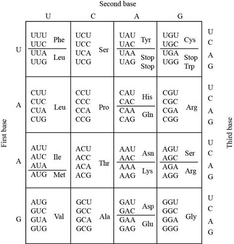
The codon table shows that the amino acid phenylalanine (Phe) is specified by onlytwo codons UUU and UUC. The amino acid serine (Ser) is specified by four codons UCA, UCC, UCA,andUCG.
The codons of serine could be developed by mutation of both codons of phenylalaninethat includes UUU and UUC. The mutation occurs at a single base position in the codon.
c.
To determine:
The position of the codon in the mRNA that must be altered by which phenylalanine is converted to isoleucine
Introduction:
The basic unit of genetic code is called a codon. The genetic code is a triplet code, in which three nucleotides encode each amino acid in a protein. The genetic code has sixty-one codons that specify the twenty amino acids. The degeneracy of genetic code means that the code is redundant and the amino acids may be specified by more than one codon.
Tryptophan and methionine are the only amino acids that are encoded by a single codon.
c.
Explanation of Solution
The codon table represents the codons and coded amino acids:

The codon table shows that the amino acid phenylalanine (Phe) is specified by only two codons UUU and UUC. The amino acid isoleucine (Ile) is specified by three codons AUU, AUC, and AUA.
The codons of isoleucine could be developed by mutation of both codons of phenylalanine that includes UUU and UUC. The mutation occurs at a single base position in the codon.
d.
To determine:
The position of the codon in the mRNA that must be altered by which proline is converted to alanine
Introduction:
The basic unit of genetic code is called a codon. The genetic code is a triplet code, in which three nucleotides encode each amino acid in a protein. The genetic code has sixty-one codons that specify the twenty amino acids. The degeneracy of genetic code means that the code is redundant and the amino acids may be specified by more than one codon.
Tryptophan and methionine are the only amino acids that are encoded by a single codon.
d.
Explanation of Solution
The codon table represents the codons and coded amino acids:

The codon table shows that the amino acid proline (Pro) is specified byfour codons CCU, CCA, CCC, and CCG. The amino acid alanine (Ala) is specified by GCU, GCG, GCC, and GCA.
The codons of alanine could be developed by mutation of all codons of proline that includes CCU, CCA, CCC, and CCG. The mutation occurs at a single base position in the codon.
e.
To determine:
The position of the codon in the mRNA that must be altered by which asparagine is converted to lysine
Introduction:
The basic unit of genetic code is called a codon. The genetic code is a triplet code, in which three nucleotides encode each amino acid in a protein. The genetic code has sixty-one codons that specify the twenty amino acids. The degeneracy of genetic code means that the code is redundant and the amino acids may be specified by more than one codon.
Tryptophan and methionine are the only amino acids that are encoded by a single codon.
e.
Explanation of Solution
The codon table represents the codons and coded amino acids:
The codon table shows that the amino acid asparagine (Asn) is specified by two codons AAU and AAC. The amino acid lysine is specified by the codons AAA and AAG.
The codons of lysine could be developed by mutation of bothcodons of asparagine that includes AAU and AAC. The mutation occurs at a single base position in the codon.
f.
To determine:
The position of the codon in the mRNA that must be altered by which isoleucine is converted to asparagine
Introduction:
The basic unit of genetic code is called a codon. The genetic code is a triplet code, in which three nucleotides encode each amino acid in a protein. The genetic code has sixty-one codons that specify the twenty amino acids. The degeneracy of genetic code means that the code is redundant and the amino acids may be specified by more than one codon.
Tryptophan and methionine are the only amino acids that are encoded by a single codon.
f.
Explanation of Solution
The codon table represents the codons and coded amino acids:
The codon table shows that the amino acid isoleucine (Ile) is specified by three codons AUU, AUC, and AUA. The amino acid asparagine is specified by the codons AAU and AAC
The codons of asparagine could be developed by mutation of only two codons of isoleucine that includes AUU and AUC. The mutation occurs at a single base position in the codon.
Want to see more full solutions like this?
Chapter 15 Solutions
Genetics: A Conceptual Approach

- Describe the principle of homeostasis.arrow_forwardExplain how the hormones of the glands listed below travel around the body to target organs and tissues : Pituitary gland Hypothalamus Thyroid Parathyroid Adrenal Pineal Pancreas(islets of langerhans) Gonads (testes and ovaries) Placentaarrow_forwardWhat are the functions of the hormones produced in the glands listed below: Pituitary gland Hypothalamus Thyroid Parathyroid Adrenal Pineal Pancreas(islets of langerhans) Gonads (testes and ovaries) Placentaarrow_forward
- Describe the hormones produced in the glands listed below: Pituitary gland Hypothalamus Thyroid Parathyroid Adrenal Pineal Pancreas(islets of langerhans) Gonads (testes and ovaries) Placentaarrow_forwardPlease help me calculate drug dosage from the following information: Patient weight: 35 pounds, so 15.9 kilograms (got this by dividing 35 pounds by 2.2 kilograms) Drug dose: 0.05mg/kg Drug concentration: 2mg/mLarrow_forwardA 25-year-old woman presents to the emergency department with a 2-day history of fever, chills, severe headache, and confusion. She recently returned from a trip to sub-Saharan Africa, where she did not take malaria prophylaxis. On examination, she is febrile (39.8°C/103.6°F) and hypotensive. Laboratory studies reveal hemoglobin of 8.0 g/dL, platelet count of 50,000/μL, and evidence of hemoglobinuria. A peripheral blood smear shows ring forms and banana-shaped gametocytes. Which of the following Plasmodium species is most likely responsible for her severe symptoms? A. Plasmodium vivax B. Plasmodium ovale C. Plasmodium malariae D. Plasmodium falciparumarrow_forward
- please fill in missing parts , thank youarrow_forwardplease draw in the answers, thank youarrow_forwarda. On this first grid, assume that the DNA and RNA templates are read left to right. DNA DNA mRNA codon tRNA anticodon polypeptide _strand strand C с A T G A U G C A TRP b. Now do this AGAIN assuming that the DNA and RNA templates are read right to left. DNA DNA strand strand C mRNA codon tRNA anticodon polypeptide 0 A T G A U G с A TRParrow_forward
 Human Anatomy & Physiology (11th Edition)BiologyISBN:9780134580999Author:Elaine N. Marieb, Katja N. HoehnPublisher:PEARSON
 Biology 2eBiologyISBN:9781947172517Author:Matthew Douglas, Jung Choi, Mary Ann ClarkPublisher:OpenStax
 Anatomy & PhysiologyBiologyISBN:9781259398629Author:McKinley, Michael P., O'loughlin, Valerie Dean, Bidle, Theresa StouterPublisher:Mcgraw Hill Education,
 Molecular Biology of the Cell (Sixth Edition)BiologyISBN:9780815344322Author:Bruce Alberts, Alexander D. Johnson, Julian Lewis, David Morgan, Martin Raff, Keith Roberts, Peter WalterPublisher:W. W. Norton & Company
 Laboratory Manual For Human Anatomy & PhysiologyBiologyISBN:9781260159363Author:Martin, Terry R., Prentice-craver, CynthiaPublisher:McGraw-Hill Publishing Co.
 Inquiry Into Life (16th Edition)BiologyISBN:9781260231700Author:Sylvia S. Mader, Michael WindelspechtPublisher:McGraw Hill Education