a.
To determine:
The position of the codon in the mRNA that must be altered by which Leucine is converted to glutamine. (
Introduction:
The basic unit of genetic code is called a codon. The genetic code is a triplet code, in which three nucleotides encode each amino acid in a protein. The genetic code has sixty-one codons that specify the twenty amino acids. The degeneracy of genetic code means that the code is redundant and the amino acids may be specified by more than one codon.
Tryptophan and methionine are the only amino acids that are encoded by a single codon.
a.
Explanation of Solution
The codon table represents the codons and coded amino acids:

The codon table shows that the amino acid Leucine (Leu) is specified by six codons CUU, CUC, CUA, CUG, UAA, and UUG. The amino acid glutamine is specified by only two codons CAA and CAG.
The codons of glutamine could be developed by mutation of two codons of Leucine that includes CUA and CUG. The mutation occurs at a single base position in the codon.
b.
To determine:
The position of the codon in the mRNA that must be altered by which phenylalanineis converted to serine
Introduction:
The basic unit of genetic code is called a codon. The genetic code is a triplet code, in which three nucleotides encode each amino acid in a protein. The genetic code has sixty-one codons that specify the twenty amino acids. The degeneracy of genetic code means that the code is redundant and the amino acids may be specified by more than one codon.
Tryptophan and methionine are the only amino acids that are encoded by a single codon.
b.
Explanation of Solution
The codon table represents the codons and coded amino acids:
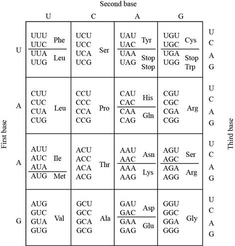
The codon table shows that the amino acid phenylalanine (Phe) is specified by onlytwo codons UUU and UUC. The amino acid serine (Ser) is specified by four codons UCA, UCC, UCA,andUCG.
The codons of serine could be developed by mutation of both codons of phenylalaninethat includes UUU and UUC. The mutation occurs at a single base position in the codon.
c.
To determine:
The position of the codon in the mRNA that must be altered by which phenylalanine is converted to isoleucine
Introduction:
The basic unit of genetic code is called a codon. The genetic code is a triplet code, in which three nucleotides encode each amino acid in a protein. The genetic code has sixty-one codons that specify the twenty amino acids. The degeneracy of genetic code means that the code is redundant and the amino acids may be specified by more than one codon.
Tryptophan and methionine are the only amino acids that are encoded by a single codon.
c.
Explanation of Solution
The codon table represents the codons and coded amino acids:

The codon table shows that the amino acid phenylalanine (Phe) is specified by only two codons UUU and UUC. The amino acid isoleucine (Ile) is specified by three codons AUU, AUC, and AUA.
The codons of isoleucine could be developed by mutation of both codons of phenylalanine that includes UUU and UUC. The mutation occurs at a single base position in the codon.
d.
To determine:
The position of the codon in the mRNA that must be altered by which proline is converted to alanine
Introduction:
The basic unit of genetic code is called a codon. The genetic code is a triplet code, in which three nucleotides encode each amino acid in a protein. The genetic code has sixty-one codons that specify the twenty amino acids. The degeneracy of genetic code means that the code is redundant and the amino acids may be specified by more than one codon.
Tryptophan and methionine are the only amino acids that are encoded by a single codon.
d.
Explanation of Solution
The codon table represents the codons and coded amino acids:

The codon table shows that the amino acid proline (Pro) is specified byfour codons CCU, CCA, CCC, and CCG. The amino acid alanine (Ala) is specified by GCU, GCG, GCC, and GCA.
The codons of alanine could be developed by mutation of all codons of proline that includes CCU, CCA, CCC, and CCG. The mutation occurs at a single base position in the codon.
e.
To determine:
The position of the codon in the mRNA that must be altered by which asparagine is converted to lysine
Introduction:
The basic unit of genetic code is called a codon. The genetic code is a triplet code, in which three nucleotides encode each amino acid in a protein. The genetic code has sixty-one codons that specify the twenty amino acids. The degeneracy of genetic code means that the code is redundant and the amino acids may be specified by more than one codon.
Tryptophan and methionine are the only amino acids that are encoded by a single codon.
e.
Explanation of Solution
The codon table represents the codons and coded amino acids:
The codon table shows that the amino acid asparagine (Asn) is specified by two codons AAU and AAC. The amino acid lysine is specified by the codons AAA and AAG.
The codons of lysine could be developed by mutation of bothcodons of asparagine that includes AAU and AAC. The mutation occurs at a single base position in the codon.
f.
To determine:
The position of the codon in the mRNA that must be altered by which isoleucine is converted to asparagine
Introduction:
The basic unit of genetic code is called a codon. The genetic code is a triplet code, in which three nucleotides encode each amino acid in a protein. The genetic code has sixty-one codons that specify the twenty amino acids. The degeneracy of genetic code means that the code is redundant and the amino acids may be specified by more than one codon.
Tryptophan and methionine are the only amino acids that are encoded by a single codon.
f.
Explanation of Solution
The codon table represents the codons and coded amino acids:
The codon table shows that the amino acid isoleucine (Ile) is specified by three codons AUU, AUC, and AUA. The amino acid asparagine is specified by the codons AAU and AAC
The codons of asparagine could be developed by mutation of only two codons of isoleucine that includes AUU and AUC. The mutation occurs at a single base position in the codon.
Want to see more full solutions like this?
Chapter 15 Solutions
Genetics: A Conceptual Approach

- Molecular Biology Question You are working to characterize a novel protein in mice. Analysis shows that high levels of the primary transcript that codes for this protein are found in tissue from the brain, muscle, liver, and pancreas. However, an antibody that recognizes the C-terminal portion of the protein indicates that the protein is present in brain, muscle, and liver, but not in the pancreas. What is the most likely explanation for this result?arrow_forwardMolecular Biology Explain/discuss how “slow stop” and “quick/fast stop” mutants wereused to identify different protein involved in DNA replication in E. coli.arrow_forwardMolecular Biology Question A gene that codes for a protein was removed from a eukaryotic cell and inserted into a prokaryotic cell. Although the gene was successfully transcribed and translated, it produced a different protein than it produced in the eukaryotic cell. What is the most likely explanation?arrow_forward
- Molecular Biology LIST three characteristics of origins of replicationarrow_forwardMolecular Biology Question Please help. Thank you For E coli DNA polymerase III, give the structure and function of the b-clamp sub-complex. Describe how the structure of this sub-complex is important for it’s function.arrow_forwardMolecular Biology LIST three characteristics of DNA Polymerasesarrow_forward
- Molecular Biology RNA polymerase core enzyme structure contains what subunits? To form holo enzyme, sigma factor is added to core. What is the name of the structure formed? Give the detailed structure of sigma factor and the function of eachdomain. Please help. Thank youarrow_forwardMolecular Biology You have a single bacterial cell whose DNA is labelled with radioactiveC14. After 5 rounds of cell division, how may cells will contain radioactive DNA? Please help. Thank youarrow_forward1. Explain the structure and properties of atoms and chemical bonds (especially how they relate to DNA and proteins). Also add some pictures.arrow_forward
- 1. In the Sentinel Cell DNA integrity is preserved through nanoscopic helicase-coordinated repair, while lipids in the membrane are fortified to resist environmental mutagens. also provide pictures for this question.arrow_forwardExplain the structure and properties of atoms and chemical bonds (especially how they relate to DNA and proteins). Also add some pictures.arrow_forwardIn the Sentinel Cell DNA integrity is preserved through nanoscopic helicase-coordinated repair, while lipids in the membrane are fortified to resist environmental mutagens. also provide pictures for this question.arrow_forward
 Human Anatomy & Physiology (11th Edition)BiologyISBN:9780134580999Author:Elaine N. Marieb, Katja N. HoehnPublisher:PEARSON
 Biology 2eBiologyISBN:9781947172517Author:Matthew Douglas, Jung Choi, Mary Ann ClarkPublisher:OpenStax
 Anatomy & PhysiologyBiologyISBN:9781259398629Author:McKinley, Michael P., O'loughlin, Valerie Dean, Bidle, Theresa StouterPublisher:Mcgraw Hill Education,
 Molecular Biology of the Cell (Sixth Edition)BiologyISBN:9780815344322Author:Bruce Alberts, Alexander D. Johnson, Julian Lewis, David Morgan, Martin Raff, Keith Roberts, Peter WalterPublisher:W. W. Norton & Company
 Laboratory Manual For Human Anatomy & PhysiologyBiologyISBN:9781260159363Author:Martin, Terry R., Prentice-craver, CynthiaPublisher:McGraw-Hill Publishing Co.
 Inquiry Into Life (16th Edition)BiologyISBN:9781260231700Author:Sylvia S. Mader, Michael WindelspechtPublisher:McGraw Hill Education