Concept explainers
Videos
The chromosome constitution number of this individual is
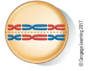
This drawing represents:
a. mitotic metaphase.
b. meiotic metaphase I.
c. meiotic metaphase II.
d. a gamete.
e. sixnonhomologous chromosomes.
Introduction:
The cell cycle consists of two main divisions known as mitosis and meiosis. Mitosis is the first division during the cell cycle in which the number of chromosomes remains same as the number of chromosomes in the parent cell; on the contrary, in meiosis, the number of chromosomes gets reduced to half.
Answer to Problem 1TYK
Correct answer:
Metaphase 1 of meiosis.
Explanation of Solution
Justification for the correct answer:
Option (b) is meiotic metaphase 1. There are two meiotic divisions after mitosis, namely, meiosis 1 and meiosis 2. During the meiosis 1, the paired chromosomes, also known as bivalent, arrange themselves on the metaphase plate. These paired chromosomes align at the middle of the plate and get attached to the meiotic spindle. The spindle is formed from the structure of the protein, which is responsible for dividing the genetic material in the cell. Hence, option (b) is correct.
Explanation for the incorrect answer:
Option (a) is metaphase in mitosis. Mitosis is the first part of the cell cycle that takes place before the meiosis. Metaphase stage in mitosis involves the arrangement of most condensed chromosomes and these chromosomes then get coiled at this stage. During the metaphase stage of meiosis, the chromosomes are not paired. So, it is an incorrect option.
Option (c) is metaphase 2 phase of meiosis 2. The chromosomes in the meiosis 2 are similar to the chromosomes found in the metaphase stage of mitosis. During the meiosis 2, the chromosomes get condensed and attach themselves to the spindles arising from the centromere present at the opposite poles. These condensed chromosomes are aligned on a metaphase plate but they are in a condensed form. So, it is an incorrect option.
Option (d) is a gamete. During fertilization fusion of one haploid gamete with another haploid gamete results in the formation of a diploid zygote. Number of chromosomes in a haploid cell is 23, two haploid cells fuse together to form a diploid cell. During meiosis number of chromosomes are reduced to 23 from 46 chromosomes. So, it is an incorrect option.
Option (e) states that number of non-homologous chromosomes is 6. Paired chromosomes are the one in which one chromosome is a homolog to another chromosome. These paired chromosomes have the same gene content and possess the same morphology as well. Non-homologous chromosomes are paired chromosomes in which both the chromosomes possess a different gene content. So, it is an incorrect option.
Hence, options (a), (c), (d), and (e) are incorrect.
Therefore, it may be concluded that in the metaphase division of meiosis 1 alignment of paired chromosomes on the metaphase plate takes place. These paired chromosomes get attached to the spindle fibers during this (metaphase 1) stage.
Want to see more full solutions like this?
Chapter 11 Solutions
Biology: The Dynamic Science (MindTap Course List)


- 1.)What cross will result in half homozygous dominant offspring and half heterozygous offspring? 2.) What cross will result in all heterozygous offspring?arrow_forward1.Steroids like testosterone and estrogen are nonpolar and large (~18 carbons). Steroids diffuse through membranes without transporters. Compare and contrast the remaining substances and circle the three substances that can diffuse through a membrane the fastest, without a transporter. Put a square around the other substance that can also diffuse through a membrane (1000x slower but also without a transporter). Molecule Steroid H+ CO₂ Glucose (C6H12O6) H₂O Na+ N₂ Size (Small/Big) Big Nonpolar/Polar/ Nonpolar lonizedarrow_forwardwhat are the answer from the bookarrow_forward
- what is lung cancer why plants removes liquid water intead water vapoursarrow_forward*Example 2: Tracing the path of an autosomal dominant trait Trait: Neurofibromatosis Forms of the trait: The dominant form is neurofibromatosis, caused by the production of an abnormal form of the protein neurofibromin. Affected individuals show spots of abnormal skin pigmentation and non-cancerous tumors that can interfere with the nervous system and cause blindness. Some tumors can convert to a cancerous form. i The recessive form is a normal protein - in other words, no neurofibromatosis.moovi A typical pedigree for a family that carries neurofibromatosis is shown below. Note that carriers are not indicated with half-colored shapes in this chart. Use the letter "N" to indicate the dominant neurofibromatosis allele, and the letter "n" for the normal allele. Nn nn nn 2 nn Nn A 3 N-arrow_forwardI want to be a super nutrition guy what u guys like recommend mearrow_forward
- Please finish the chart at the bottom. Some of the answers have been filled in.arrow_forward9. Aerobic respiration of one lipid molecule. The lipid is composed of one glycerol molecule connected to two fatty acid tails. One fatty acid is 12 carbons long and the other fatty acid is 18 carbons long in the figure below. Use the information below to determine how much ATP will be produced from the glycerol part of the lipid. Then, in part B, determine how much ATP is produced from the 2 fatty acids of the lipid. Finally put the NADH and ATP yields together from the glycerol and fatty acids (part A and B) to determine your total number of ATP produced per lipid. Assume no other carbon source is available. 18 carbons fatty acids 12 carbons 9 glycerol A. Glycerol is broken down to glyceraldehyde 3-phosphate, a glycolysis intermediate via the following pathway shown in the figure below. Notice this process costs one ATP but generates one FADH2. Continue generating ATP with glyceraldehyde-3-phosphate using the standard pathway and aerobic respiration. glycerol glycerol-3- phosphate…arrow_forwardNormal dive (for diving humans) normal breathing dive normal breathing Oz level CO2 level urgent need to breathe Oz blackout zone high CO2 triggers breathing 6. This diagram shows rates of oxygen depletion and carbon dioxide accumulation in the blood in relation to the levels needed to maintain consciousness and trigger the urgent need to breathe in diving humans. How might the location and slope of the O₂ line differ for diving marine mammals such as whales and dolphins? • How might the location and slope of the CO₂ line differ for diving marine mammals such as whales and dolphins? • • Draw in predicted lines for O2 and CO2, based on your reasoning above. How might the location of the Urgent Need to Breathe line and the O2 Blackout Zone line differ for diving marine mammals? What physiological mechanisms account for each of these differences, resulting in the ability of marine mammals to stay submerged for long periods of time?arrow_forward
- foraging/diet type teeth tongue stomach intestines cecum Insectivory numerous, spiky, incisors procumbentExample: moleExample: shrew -- simple short mostly lacking Myrmecophagy absent or reduced in numbers, peg-likeExample: tamandua anteater extremely long simple, often roughened short small or lacking Terrestrial carnivory sharp incisors; long, conical canines; often carnassial cheek teeth; may have crushing molarsExample: dog -- simple short small Aquatic carnivory homodont, spiky, numerousExample: common dolphin -- simple or multichambered (cetaceans only) variable small or absent Sanguinivory very sharp upper incisors; reduced cheek teethExample: vampire bat grooved tubular, highly extensible long small or lacking Herbivory (except nectivores) incisors robust or absent; canines reduced or absent; diastema; cheek teeth enlarged with complex occlusal surfacesExample: beaver -- simple (hindgut fermenters) or multichambered (ruminants) long large Filter feeding none…arrow_forward3. Shown below is the dental formula and digestive tract anatomy of three mammalian species (A, B, and C). What kind of diet would you expect each species to have? Support your answers with what you can infer from the dental formula and what you can see in the diagram. Broadly speaking, what accounts for the differences? Species A 3/3, 1/1, 4/4, 3/3 པར『ན་ cm 30 Species B 4/3, 1/1, 2/2, 4/4 cm 10 Species C 0/4, 0/0,3/3, 3/3 020arrow_forward3. Shown below is the dental formula and digestive tract anatomy of three mammalian species (A, B, and C). What kind of diet would you expect each species to have? Support your answers with what you can infer from the dental formula and what you can see in the diagram. Broadly speaking, what accounts for the differences? Species A 3/3, 1/1, 4/4, 3/3 cm 30 Species B 0/4, 0/0, 3/3, 3/3 cm 10 Species C 4/3, 1/1, 2/2, 4/4 E 0 cm 20 AILarrow_forward
 Biology: The Dynamic Science (MindTap Course List)BiologyISBN:9781305389892Author:Peter J. Russell, Paul E. Hertz, Beverly McMillanPublisher:Cengage Learning
 Biology Today and Tomorrow without Physiology (Mi...BiologyISBN:9781305117396Author:Cecie Starr, Christine Evers, Lisa StarrPublisher:Cengage Learning
 Concepts of BiologyBiologyISBN:9781938168116Author:Samantha Fowler, Rebecca Roush, James WisePublisher:OpenStax College
 Biology 2eBiologyISBN:9781947172517Author:Matthew Douglas, Jung Choi, Mary Ann ClarkPublisher:OpenStax
 Human Heredity: Principles and Issues (MindTap Co...BiologyISBN:9781305251052Author:Michael CummingsPublisher:Cengage Learning