
Concept explainers
Videos
To label: The features of the long bone of an adult.
Introduction: The long bones are typically found in the upper and lower extremities such as thighs and arms. They are the hard and dense bones that provide structure, strength, and mobility to the body. The gross anatomy of long bones includes epiphysis, diaphysis, endosteum, articular cartilage, medullary cavity, compact bone, spongy bone, metaphysis, and periosteum.
Answer to Problem 1.1BGL
Pictorial representation:
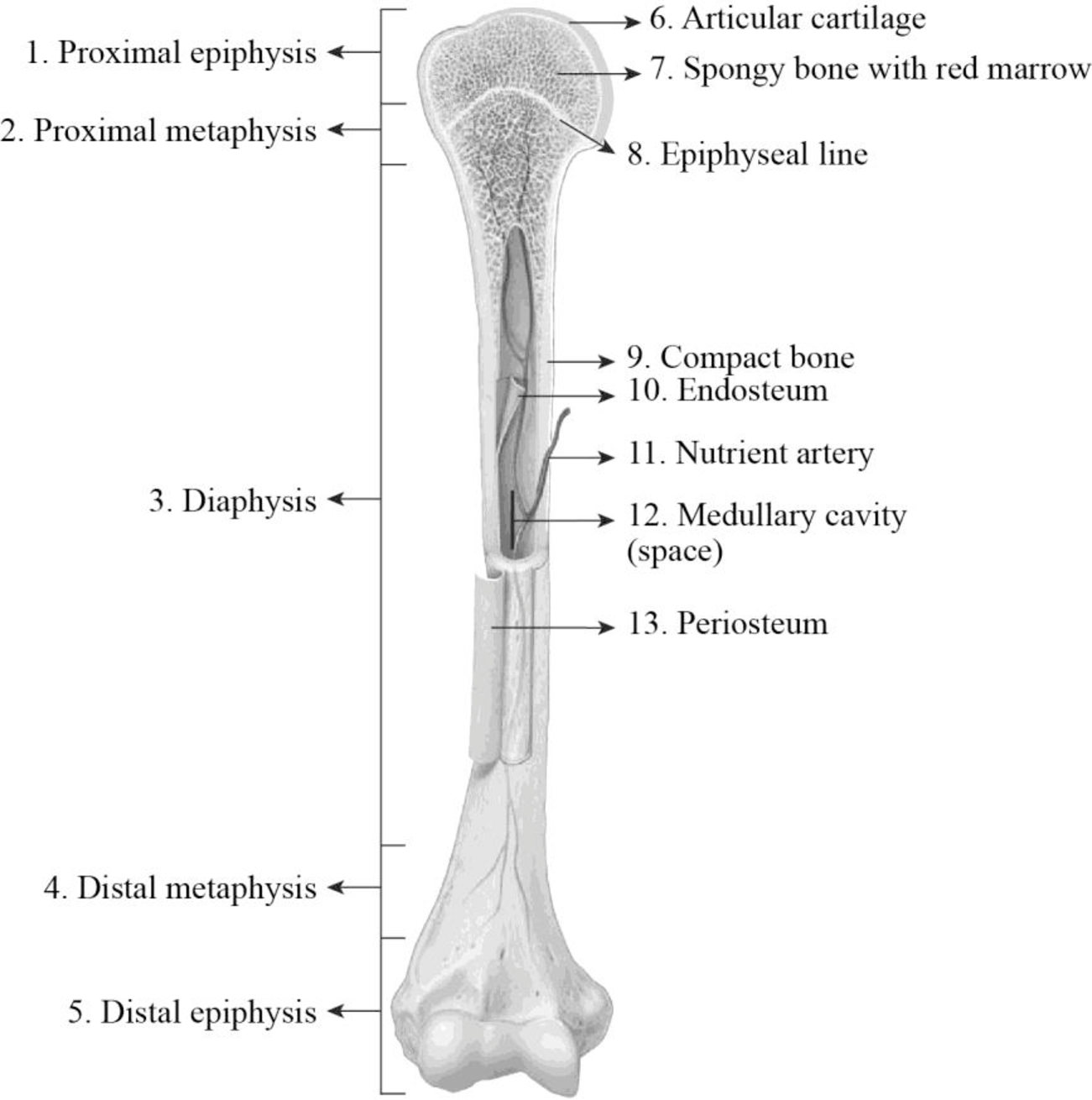
Fig1: Features of the long bone of an adult.
Explanation of Solution
1. Proximal epiphysis: The area of the bone that is covered with the cartilage is the epiphysis. The wider section of the proximal and distal end of the long bone is termed as epiphysis. It is made up of spongy cancellous bone and it is enclosed by a thin layer of compact bone. The spongy spaces of the epiphyses are filled with red bone marrow. The closer end of the long bone is referred to as distal epiphysis.
2. Proximal metaphysis: In adults, the area where the diaphysis and epiphysis join together is referred to as metaphysis. The metaphysis that is located closer to the center of the body is termed as proximal metaphysis.
3. Diaphysis: The diaphysis is the layer that is made up of very thin layer of spongy bone and thick layer of compact bone. The diaphysis is the mid-section of the long bone that is generally termed as shaft. It is made up of the cortical bone and usually contains fat (adipose tissue) and bone marrow. It is a tubular part that is composed of the thick layers of compact bones and the thin layer of spongy bones. The compact bones cover the exterior part, and the spongy bone lines the interior part of the diaphysis.
4. Distal metaphysis: In adults, the area where diaphysis and epiphysis join together is referred to as metaphysis. The metaphysis that is located farther from the center of the body is termed as distal metaphysis.
5. Distal epiphysis: The area of the bone that is covered with the cartilage is the epiphysis. The wider section of the proximal and distal end of the long bone is termed as epiphysis. It is made up of spongy cancellous bone and it is covered by a thin layer of compact bone. The spongy spaces of the epiphyses are filled with red bone marrow. The far end of the long bone is referred to as distal epiphysis.
6. Articular cartilage: It is a thin layer of specialized connective tissue that has unique viscoelastic properties. The main function of this articular cartilage is to provide a smooth and lubricate surface to the bones. It provides low-friction articulation and provides the transmission of loads to the interior subchondral bone. The articular cartilage is a type of hyaline cartilage. It is a translucent, grayish-white tissue that is made up of collagen proteins. It provides flexibility and support to the bones.
7. Spongy bone with red marrow: The spongy bones are also referred to as trabecular bones. This type of bones is highly porous and vascularised, which is usually located at the end of the bones. Trabeculae are the flat plates containing a lattice-like network of thin bony columns in the spongy bones. This is because the spongy bones do not possess osteons as they have large spaces, and the osteons were replaced by the trabeculae. The spaces between trabeculae are filled with red bone marrow.
8. Epiphyseal line: Generally, the growth plates are situated at the widened part of the metaphyses and at the end of the epiphyses. In a developing bone, the metaphyses possess a layer of hyaline cartilage which is referred to as an epiphyseal plate. The division in this cartilage allows for the lengthy growth of the bone. In adults, the bone growth stops when the epiphyseal plate cartilage becomes ossified thereby forming the epiphyseal line.
9. Compact bone: Compact bones are the denser materials that form the hard structure of the skeleton. The compact bones are extremely tough, heavy, and are stacked or formed in layers. It is a type of osseous tissue that is also referred to as cortical bones. They are the dense form of the bone that is made up of osteons, which makes the structure to it. The compact bones form the hard surface layer to the bones. The compact bones are made up of microscopical structural unit called an osteon or haversian system.
10. Endosteum: It is the thin vascular membrane of connective tissue which lines the medullary cavity of long bones. It helps in maintaining the integrity, shape, and mechanical properties of the bone. It provides protection and coverage to the inner parts of the bone and is also involved in the process of healing.
11. Nutrient artery: It is a large artery that enters into the compact bone closer to the middle of the diaphysis. It branches immediately into distal and proximal portions and is involved in supplying blood to the inner layers of the spongy bone, compact bone, and red marrow.
12. Medullary cavity: The cavity of the bone that is filled with bone marrow is referred to as a medullary cavity. In adults, this cavity is filled with yellow bone marrow. It is a fatty bone marrow that does not involve in the production of blood cells and it is stored in the medullary cavity. It is the innermost cavity of the bone shafts that possess both red bone marrow at a younger age and yellow bone marrow in adults.
13. Periosteum: The periosteum is a dense layer of vascular connective tissues. It envelops the surface of the bone except for the surface of joints. It protects the bone and serves as a channel for the supply of blood and nutrients to the bone tissue. The periosteum consists of a cellular inner layer and fibrous outer layer. It plays an important role in the growth and repair of the bony tissues.
Want to see more full solutions like this?
Chapter 8 Solutions
Laboratory Manual for Anatomy and Physiology, 6e WileyPLUS (next generation) + Loose-leaf


- View History Bookmarks Window Help Quarter cements ents ons (17) YouTube Which amino acids would you expect to find marked on the alpha helix? canvas.ucsc.edu ucsc Complaint and Grievance Process - Academic Personnel pach orations | | | | | | | | | | | | | | | | 000000 000000000 | | | | | | | | | | | | | | | | | | | | | | 00000000 scope vious De 48 12.415 KATPM FEB 3 F1 F2 80 F3 a F4 F5 2 # 3 $ 85 % tv N A の Mon Feb 3 10:24 PM Lipid bilayer Submit Assignment Next > ZOOM < Å DII 8 བ བ F6 16 F7 F8 F9 F10 34 F11 F12 & * ( 6 7 8 9 0 + 11 WERTY U { 0 } P deletearrow_forwardDifferent species or organisms research for ecologyarrow_forwardWhat is the result of the following gram stain: positive ○ capsulated ○ acid-fast ○ negativearrow_forward
- What type of stain is the image below: capsule stain endospore stain gram stain negative stain ASM MicrobeLibrary.org Keplingerarrow_forwardWhat is the result of the acid-fast stain below: Stock Images by Getty Images by Getty Images by Getty Images by Getty Image Getty Images St Soy Getty Images by Getty Images by Getty Images Joy Getty encapsulated O endosporulating negative ○ positivearrow_forwardYou have a stock vial of diligence 75mg in 3ml and need to draw up a dose of 50mg for your patient.how many mls should you draw up to give this dosearrow_forward
- You are recquired to administer 150mg hydrocortisone intravenously,how many mls should you give?(stock =hydrocortisone 100mg in 2mls)arrow_forwardIf someone was working with a 50 MBq F-18 source, what would be the internal and external dose consequences?arrow_forwardWe will be starting a group project next week where you and your group will research and ultimately present on a current research article related to the biology of a pathogen that infects humans. The article could be about the pathogen itself, the disease process related to the pathogen, the immune response to the pathogen, vaccines or treatments that affect the pathogen, or other biology-related study about the pathogen. I recommend that you choose a pathogen that is currently interesting to researchers, so that you will be able to find plenty of articles about it. Avoid choosing a historical disease that no longer circulates. List 3 possible pathogens or diseases that you might want to do for your group project.arrow_forward
- not use ai pleasearrow_forwardDNK dagi nukleotidlar va undan sintezlangan oqsildagi peptid boglar farqi 901 taga teng bo'lib undagi A jami H boglardan 6,5 marta kam bo'lsa DNK dagi jami H bog‘lar sonini topingarrow_forwardOne of the ways for a cell to generate ATP is through the oxidative phosphorylation. In oxidative phosphorylation 3 ATP are produced from every one NADH molecule. In respiration, every glucose molecule produces 10 NADH molecules. If a cell is growing on 5 glucose molecules, how much ATP can be produced using oxidative phosphorylation/aerobic respiration?arrow_forward
 Human Anatomy & Physiology (11th Edition)BiologyISBN:9780134580999Author:Elaine N. Marieb, Katja N. HoehnPublisher:PEARSON
 Biology 2eBiologyISBN:9781947172517Author:Matthew Douglas, Jung Choi, Mary Ann ClarkPublisher:OpenStax
 Anatomy & PhysiologyBiologyISBN:9781259398629Author:McKinley, Michael P., O'loughlin, Valerie Dean, Bidle, Theresa StouterPublisher:Mcgraw Hill Education,
 Molecular Biology of the Cell (Sixth Edition)BiologyISBN:9780815344322Author:Bruce Alberts, Alexander D. Johnson, Julian Lewis, David Morgan, Martin Raff, Keith Roberts, Peter WalterPublisher:W. W. Norton & Company
 Laboratory Manual For Human Anatomy & PhysiologyBiologyISBN:9781260159363Author:Martin, Terry R., Prentice-craver, CynthiaPublisher:McGraw-Hill Publishing Co.
 Inquiry Into Life (16th Edition)BiologyISBN:9781260231700Author:Sylvia S. Mader, Michael WindelspechtPublisher:McGraw Hill Education