
Concept explainers
Videos
To label: The features of the long bone of an adult.
Introduction: The long bones are typically found in the upper and lower extremities such as thighs and arms. They are the hard and dense bones that provide structure, strength, and mobility to the body. The gross anatomy of long bones includes epiphysis, diaphysis, endosteum, articular cartilage, medullary cavity, compact bone, spongy bone, metaphysis, and periosteum.
Answer to Problem 1.1BGL
Pictorial representation:
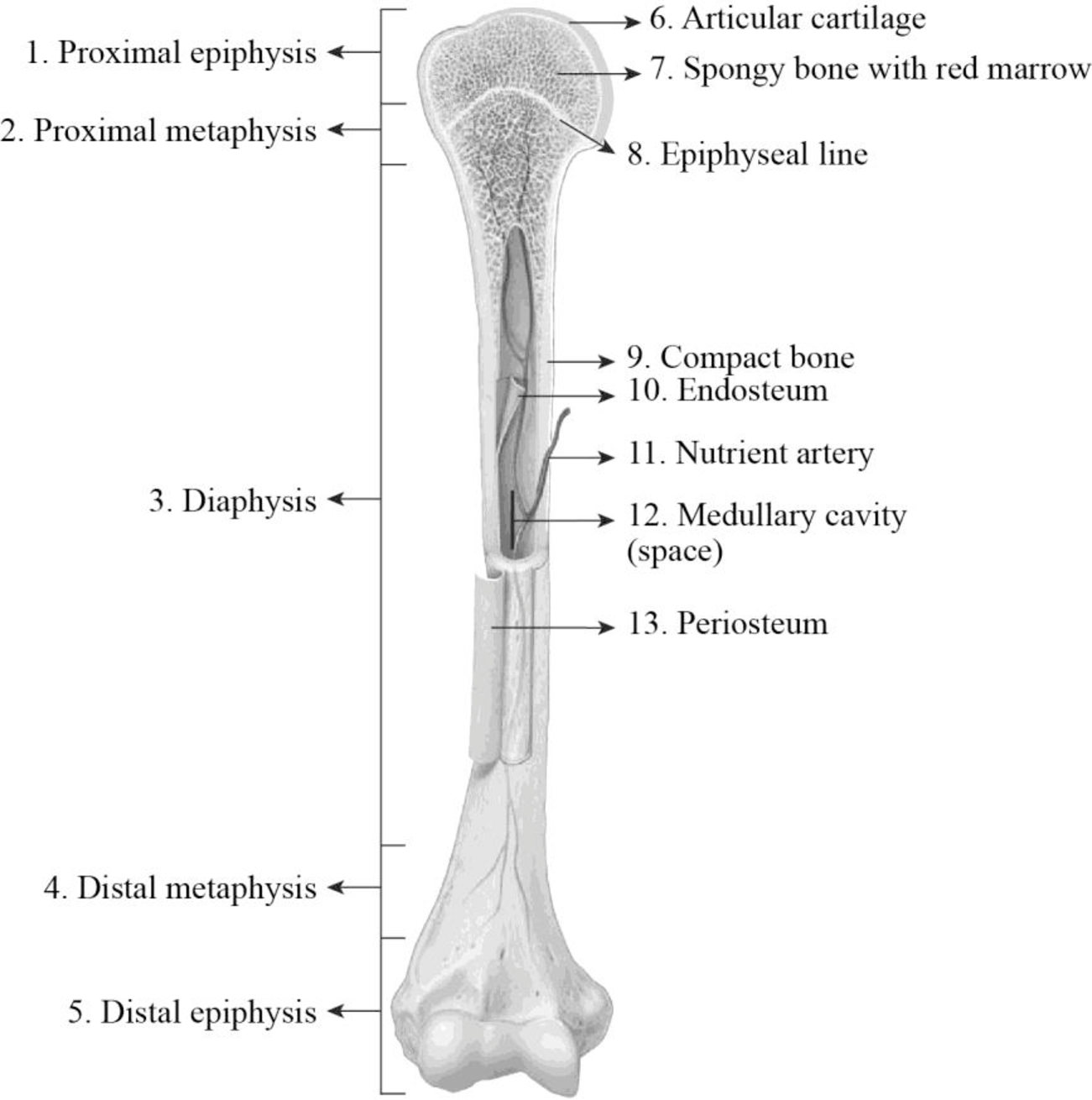
Fig1: Features of the long bone of an adult.
Explanation of Solution
1. Proximal epiphysis: The area of the bone that is covered with the cartilage is the epiphysis. The wider section of the proximal and distal end of the long bone is termed as epiphysis. It is made up of spongy cancellous bone and it is enclosed by a thin layer of compact bone. The spongy spaces of the epiphyses are filled with red bone marrow. The closer end of the long bone is referred to as distal epiphysis.
2. Proximal metaphysis: In adults, the area where the diaphysis and epiphysis join together is referred to as metaphysis. The metaphysis that is located closer to the center of the body is termed as proximal metaphysis.
3. Diaphysis: The diaphysis is the layer that is made up of very thin layer of spongy bone and thick layer of compact bone. The diaphysis is the mid-section of the long bone that is generally termed as shaft. It is made up of the cortical bone and usually contains fat (adipose tissue) and bone marrow. It is a tubular part that is composed of the thick layers of compact bones and the thin layer of spongy bones. The compact bones cover the exterior part, and the spongy bone lines the interior part of the diaphysis.
4. Distal metaphysis: In adults, the area where diaphysis and epiphysis join together is referred to as metaphysis. The metaphysis that is located farther from the center of the body is termed as distal metaphysis.
5. Distal epiphysis: The area of the bone that is covered with the cartilage is the epiphysis. The wider section of the proximal and distal end of the long bone is termed as epiphysis. It is made up of spongy cancellous bone and it is covered by a thin layer of compact bone. The spongy spaces of the epiphyses are filled with red bone marrow. The far end of the long bone is referred to as distal epiphysis.
6. Articular cartilage: It is a thin layer of specialized connective tissue that has unique viscoelastic properties. The main function of this articular cartilage is to provide a smooth and lubricate surface to the bones. It provides low-friction articulation and provides the transmission of loads to the interior subchondral bone. The articular cartilage is a type of hyaline cartilage. It is a translucent, grayish-white tissue that is made up of collagen proteins. It provides flexibility and support to the bones.
7. Spongy bone with red marrow: The spongy bones are also referred to as trabecular bones. This type of bones is highly porous and vascularised, which is usually located at the end of the bones. Trabeculae are the flat plates containing a lattice-like network of thin bony columns in the spongy bones. This is because the spongy bones do not possess osteons as they have large spaces, and the osteons were replaced by the trabeculae. The spaces between trabeculae are filled with red bone marrow.
8. Epiphyseal line: Generally, the growth plates are situated at the widened part of the metaphyses and at the end of the epiphyses. In a developing bone, the metaphyses possess a layer of hyaline cartilage which is referred to as an epiphyseal plate. The division in this cartilage allows for the lengthy growth of the bone. In adults, the bone growth stops when the epiphyseal plate cartilage becomes ossified thereby forming the epiphyseal line.
9. Compact bone: Compact bones are the denser materials that form the hard structure of the skeleton. The compact bones are extremely tough, heavy, and are stacked or formed in layers. It is a type of osseous tissue that is also referred to as cortical bones. They are the dense form of the bone that is made up of osteons, which makes the structure to it. The compact bones form the hard surface layer to the bones. The compact bones are made up of microscopical structural unit called an osteon or haversian system.
10. Endosteum: It is the thin vascular membrane of connective tissue which lines the medullary cavity of long bones. It helps in maintaining the integrity, shape, and mechanical properties of the bone. It provides protection and coverage to the inner parts of the bone and is also involved in the process of healing.
11. Nutrient artery: It is a large artery that enters into the compact bone closer to the middle of the diaphysis. It branches immediately into distal and proximal portions and is involved in supplying blood to the inner layers of the spongy bone, compact bone, and red marrow.
12. Medullary cavity: The cavity of the bone that is filled with bone marrow is referred to as a medullary cavity. In adults, this cavity is filled with yellow bone marrow. It is a fatty bone marrow that does not involve in the production of blood cells and it is stored in the medullary cavity. It is the innermost cavity of the bone shafts that possess both red bone marrow at a younger age and yellow bone marrow in adults.
13. Periosteum: The periosteum is a dense layer of vascular connective tissues. It envelops the surface of the bone except for the surface of joints. It protects the bone and serves as a channel for the supply of blood and nutrients to the bone tissue. The periosteum consists of a cellular inner layer and fibrous outer layer. It plays an important role in the growth and repair of the bony tissues.
Want to see more full solutions like this?
Chapter 8 Solutions
Laboratory Manual for Anatomy and Physiology, 6e Loose-Leaf Print Companion with WileyPLUS Blackboard Card Set


- Transcription and Translation 1. What is the main function of transcription and translation? (2 marks) 2. How is transcription different in eukaryotic and prokaryotic cells? (2 marks) 3. Explain the difference between pre-mRNA and post-transcript mRNA. (2 marks) 4. What is the function of the following: (4 marks) i. the cap ii. spliceosome iii. Poly A tail iv. termination sequence 5. What are advantages to the wobble feature of the genetic code? (2 marks) 6. Explain the difference between the: (3 marks) i. A site & P site ii. codon & anticodon iii. gene expression and gene regulation 7. Explain how the stop codon allows for termination. (1 mark) 8. In your own words, summarize the process of translation. (2 marks)arrow_forwardIn this activity you will research performance enhancers that affect the endocrine system or nervous system. You will submit a 1 page paper on one performance enhancer of your choice. Be sure to include: the specific reason for use the alleged results on improving performance how it works how it affect homeostasis and improves performance any side-effects of this substancearrow_forwardNeurons and Reflexes 1. Describe the function of the: a) dendrite b) axon c) cell body d) myelin sheath e) nodes of Ranvier f) Schwann cells g) motor neuron, interneuron and sensory neuron 2. List some simple reflexes. Explain why babies are born with simple reflexes. What are they and why are they necessary. 3. Explain why you only feel pain after a few seconds when you touch something very hot but you have already pulled your hand away. 4. What part of the brain receives sensory information? What part of the brain directs you to move your hand away? 5. In your own words describe how the axon fires.arrow_forward
- Mutations Here is your template DNA strand: CTT TTA TAG TAG ATA CCA CAA AGG 1. Write out the complementary mRNA that matches the DNA above. 2. Write the anticodons and the amino acid sequence. 3. Change the nucleotide in position #15 to C. 4. What type of mutation is this? 5. Repeat steps 1 & 2. 6. How has this change affected the amino acid sequence? 7. Now remove nucleotides 13 through 15. 8. Repeat steps 1 & 2. 9. What type of mutation is this? 0. Do all mutations result in a change in the amino acid sequence? 1. Are all mutations considered bad? 2. The above sequence codes for a genetic disorder called cystic fibrosis (CF). 3. When A is changed to G in position #15, the person does not have CF. When T is changed to C in position #14, the person has the disorder. How could this have originated?arrow_forwardhoose a scientist(s) and research their contribution to our derstanding of DNA structure or replication. Write a one page port and include: their research where they studied and the time period in which they worked their experiments and results the contribution to our understanding of DNA cientists Watson & Crickarrow_forwardhoose a scientist(s) and research their contribution to our derstanding of DNA structure or replication. Write a one page port and include: their research where they studied and the time period in which they worked their experiments and results the contribution to our understanding of DNA cientists Watson & Crickarrow_forward
- 7. Aerobic respiration of a protein that breaks down into 12 molecules of malic acid. Assume there is no other carbon source and no acetyl-CoA. NADH FADH2 OP ATP SLP ATP Total ATP Show your work using dimensional analysis here: 3arrow_forwardFor each of the following problems calculate the following: (Week 6-3 Video with 6-1 and 6-2) Consult the total catabolic pathways on the last page as a reference for the following questions. A. How much NADH and FADH2 is produced and fed into the electron transport chain (If any)? B. How much ATP is made from oxidative phosphorylation (OP), if any? Feed the NADH and FADH2 into the electron transport chain: 3ATP/NADH, 2ATP/FADH2 C. How much ATP is made by substrate level phosphorylation (SLP)? D. How much total ATP is made? Add the SLP and OP together. 1. Aerobic respiration using 0.5 mole of glucose? NADH FADH2 OP ATP SLP ATP Total ATP Show your work using dimensional analysis here:arrow_forwardAerobic respiration of one lipid molecule. The lipid is composed of one glycerol molecule connected to two fatty acid tails. One fatty acid is 12 carbons long and the other fatty acid is 18 carbons long in the figure below. Use the information below to determine how much ATP will be produced from the glycerol part of the lipid. Then, in part B, determine how much ATP is produced from the 2 fatty acids of the lipid. Finally put the NADH and ATP yields together from the glycerol and fatty acids (part A and B) to determine your total number of ATP produced per lipid. Assume no other carbon source is available. 18 carbons fatty acids 12 carbons glycerol . Glycerol is broken down to glyceraldehyde 3-phosphate, a glycolysis intermediate via the following pathway shown in the figure below. Notice this process costs one ATP but generates one FADH2. Continue generating ATP with glyceraldehyde-3-phosphate using the standard pathway and aerobic respiration. glycerol glycerol-3- phosphate…arrow_forward
- Don't copy the other answerarrow_forward4. Aerobic respiration of 5 mM acetate solution. Assume no other carbon source and that acetate is equivalent to acetyl-CoA. NADH FADH2 OP ATP SLP ATP Total ATP Show your work using dimensional analysis here: 5. Aerobic respiration of 2 mM alpha-ketoglutaric acid solution. Assume no other carbon source. NADH FADH2 OP ATP Show your work using dimensional analysis here: SLP ATP Total ATParrow_forwardBiology You’re going to analyze 5 ul of your PCR product(out of 50 ul) on the gel. How much of 6X DNAloading buffer (dye) are you going to mix with yourPCR product to make final 1X concentration ofloading buffer in the PCR product-loading buffermixture?arrow_forward
 Human Anatomy & Physiology (11th Edition)BiologyISBN:9780134580999Author:Elaine N. Marieb, Katja N. HoehnPublisher:PEARSON
 Biology 2eBiologyISBN:9781947172517Author:Matthew Douglas, Jung Choi, Mary Ann ClarkPublisher:OpenStax
 Anatomy & PhysiologyBiologyISBN:9781259398629Author:McKinley, Michael P., O'loughlin, Valerie Dean, Bidle, Theresa StouterPublisher:Mcgraw Hill Education,
 Molecular Biology of the Cell (Sixth Edition)BiologyISBN:9780815344322Author:Bruce Alberts, Alexander D. Johnson, Julian Lewis, David Morgan, Martin Raff, Keith Roberts, Peter WalterPublisher:W. W. Norton & Company
 Laboratory Manual For Human Anatomy & PhysiologyBiologyISBN:9781260159363Author:Martin, Terry R., Prentice-craver, CynthiaPublisher:McGraw-Hill Publishing Co.
 Inquiry Into Life (16th Edition)BiologyISBN:9781260231700Author:Sylvia S. Mader, Michael WindelspechtPublisher:McGraw Hill Education