BIO THE DNA SPRING . A DNA molecule, with its double-helix structure, can in some situations behave like a spring. Measuring the force required to stretch single DNA molecules under various conditions can provide information about the biophysical properties of DNA. A technique for measuring the stretching force makes use of a very small cantilever, which consists of a beam that is supported at one end and is free to move at the other end. like a tiny diving board. The cantilever is constructed so that it obeys Hooke’s law—that is. the displacement of its free end is proportional to the force applied to it. Because different cantilevers have different force constants, the cantilever's response must first be calibrated by applying a known force and determining the resulting deflection of the cantilever. Then one end of a DNA molecule is attached to the free end of the cantilever, and the other end of the DNA molecule is attached to a small stage that can be moved away from the cantilever, stretching the DNA. The stretched DNA pulls on the cantilever, deflecting the end of the cantilever very slightly. The measured deflection is then used to determine the force on the DNA molecule. 7.83 Based on Fig. P7.82, how much elastic potential energy is stored in the DNA when it is stretched 50 nm? (a) 2.5 × 10 −19 J; (b) 1.2 × 10 −19 J; (c) 5.0 × 10 −12 J; (d)2.5 × 10 −12 J.
BIO THE DNA SPRING . A DNA molecule, with its double-helix structure, can in some situations behave like a spring. Measuring the force required to stretch single DNA molecules under various conditions can provide information about the biophysical properties of DNA. A technique for measuring the stretching force makes use of a very small cantilever, which consists of a beam that is supported at one end and is free to move at the other end. like a tiny diving board. The cantilever is constructed so that it obeys Hooke’s law—that is. the displacement of its free end is proportional to the force applied to it. Because different cantilevers have different force constants, the cantilever's response must first be calibrated by applying a known force and determining the resulting deflection of the cantilever. Then one end of a DNA molecule is attached to the free end of the cantilever, and the other end of the DNA molecule is attached to a small stage that can be moved away from the cantilever, stretching the DNA. The stretched DNA pulls on the cantilever, deflecting the end of the cantilever very slightly. The measured deflection is then used to determine the force on the DNA molecule. 7.83 Based on Fig. P7.82, how much elastic potential energy is stored in the DNA when it is stretched 50 nm? (a) 2.5 × 10 −19 J; (b) 1.2 × 10 −19 J; (c) 5.0 × 10 −12 J; (d)2.5 × 10 −12 J.
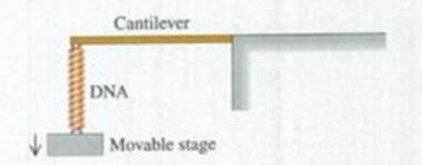
BIO THE DNA SPRING. A DNA molecule, with its double-helix structure, can in some situations behave like a spring. Measuring the force required to stretch single DNA molecules under various conditions can provide information about the biophysical properties of DNA. A technique for measuring the stretching force makes use of a very small cantilever, which consists of a beam that is supported at one end and is free to move at the other end. like a tiny diving board. The cantilever is constructed so that it obeys Hooke’s law—that is. the displacement of its free end is proportional to the force applied to it. Because different cantilevers have different force constants, the cantilever's response must first be calibrated by applying a known force and determining the resulting deflection of the cantilever. Then one end of a DNA molecule is attached to the free end of the cantilever, and the other end of the DNA molecule is attached to a small stage that can be moved away from the cantilever, stretching the DNA. The stretched DNA pulls on the cantilever, deflecting the end of the cantilever very slightly. The measured deflection is then used to determine the force on the DNA molecule.
7.83 Based on Fig. P7.82, how much elastic potential energy is stored in the DNA when it is stretched 50 nm? (a) 2.5 × 10⁻¹⁹ J; (b) 1.2 × 10⁻¹⁹ J; (c) 5.0 × 10⁻¹² J; (d)2.5 × 10⁻¹² J.
The force of the quadriceps (Fq) and force of the patellar tendon (Fp) is identical (i.e., 1000 N each). In the figure below angle in blue is Θ and the in green is half Θ (i.e., Θ/2). A) Calculate the patellar reaction force (i.e., R resultant vector is the sum of the horizontal component of the quadriceps and patellar tendon force) at the following joint angles: you need to provide a diagram showing the vector and its components for each part. a1) Θ = 160 degrees, a2) Θ = 90 degrees. NOTE: USE ONLY TRIGNOMETRIC FUNCTIONS (SIN/TAN/COS, NO LAW OF COSINES, NO COMPLICATED ALGEBRAIC EQUATIONS OR ANYTHING ELSE, ETC. Question A has 2 parts!
No chatgpt pls will upvote
Chapter 7 Solutions
Mastering Physics with Pearson eText -- Standalone Access Card -- for University Physics with Modern Physics (14th Edition)
Need a deep-dive on the concept behind this application? Look no further. Learn more about this topic, physics and related others by exploring similar questions and additional content below.
Work and Energy - Physics 101 / AP Physics 1 Review with Dianna Cowern; Author: Physics Girl;https://www.youtube.com/watch?v=rKwK06stPS8;License: Standard YouTube License, CC-BY


 Physics for Scientists and Engineers: Foundations...PhysicsISBN:9781133939146Author:Katz, Debora M.Publisher:Cengage Learning
 Principles of Physics: A Calculus-Based TextPhysicsISBN:9781133104261Author:Raymond A. Serway, John W. JewettPublisher:Cengage Learning
 Physics for Scientists and Engineers with Modern ...PhysicsISBN:9781337553292Author:Raymond A. Serway, John W. JewettPublisher:Cengage Learning
 Physics for Scientists and EngineersPhysicsISBN:9781337553278Author:Raymond A. Serway, John W. JewettPublisher:Cengage Learning
 Physics for Scientists and Engineers, Technology ...PhysicsISBN:9781305116399Author:Raymond A. Serway, John W. JewettPublisher:Cengage Learning
 College PhysicsPhysicsISBN:9781938168000Author:Paul Peter Urone, Roger HinrichsPublisher:OpenStax College