Concept explainers
Heat lamps are commonly used to maintain foods at about 50°C for as long as 12 hours in cafeteria serving lines. The following experiment was conducted to determine whether this practice poses a potential health hazard.
Beef cubes were surface-inoculated with 500,000 bacterial cells and incubated at 43–53°C to establish temperature limits for bacterial growth. The following results were obtained from heterotrophic plate counts performed on beef cubes at 6 and 12 hours after inoculation:
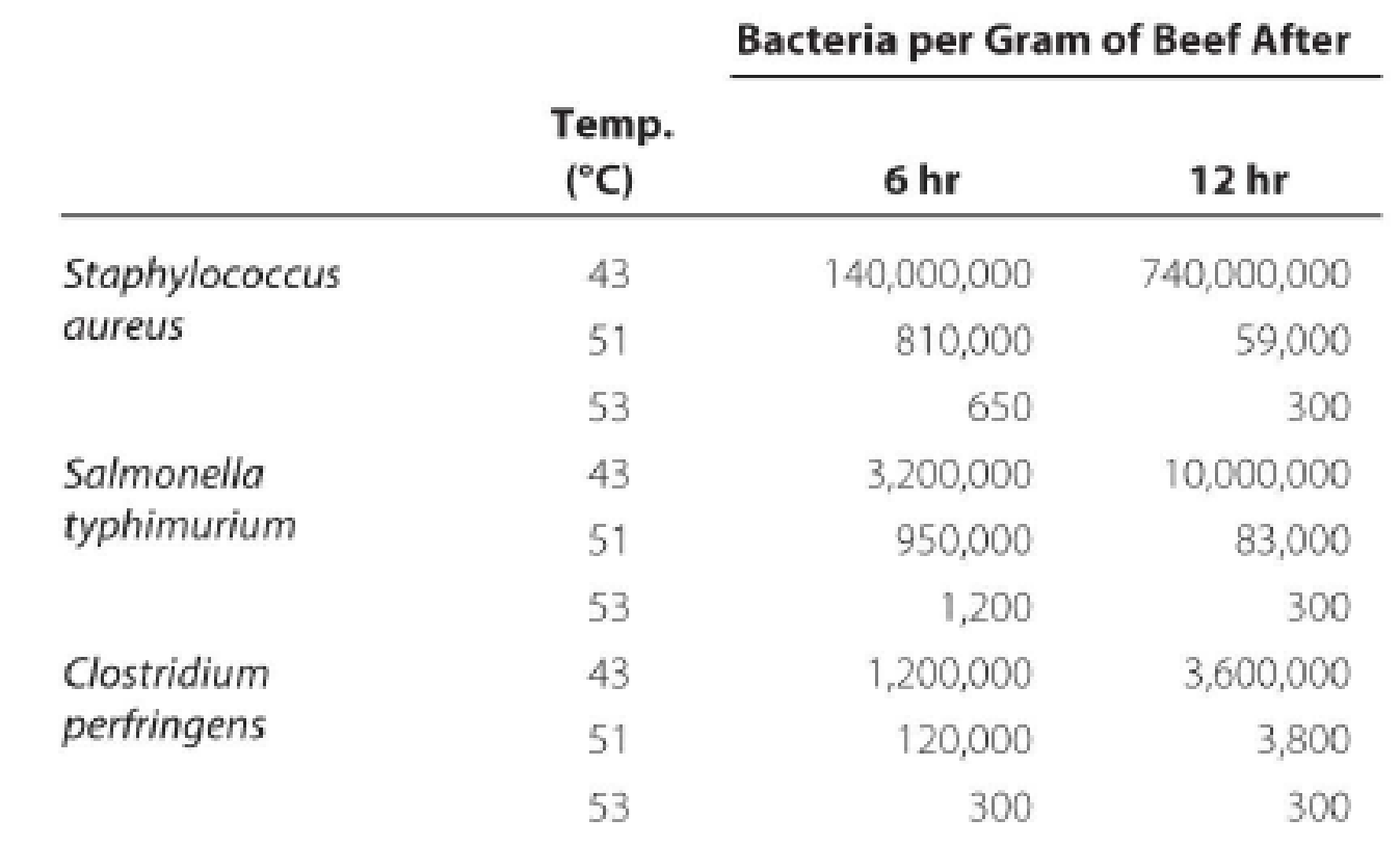
Draw the growth curves for each organism. What holding temperature would you recommend? Assuming that cooking kills bacteria in foods, how could these bacteria contaminate the cooked foods? What disease does each organism cause? (Hint: See Chapter 25.)
Want to see the full answer?
Check out a sample textbook solutionChapter 6 Solutions
MICROBIOLOGY-ACCESS >CUSTOM<
Additional Science Textbook Solutions
Microbiology with Diseases by Body System (4th Edition)
Concepts of Genetics (11th Edition)
Human Anatomy & Physiology (11th Edition)
Campbell Essential Biology (6th Edition) - standalone book
Genetic Analysis: An Integrated Approach (3rd Edition)
Laboratory Experiments in Microbiology (12th Edition) (What's New in Microbiology)
- Using one of the graphs obtained with the medium II, name the different phases of the bacterial growth and indicate the starting and ending time points of these phases as well as the physiological characteristics of the bacteria during each phasearrow_forwardYou spread 0.1 mL volume of a 10^(-6) dilution onto a nutrient agar plate. After 24 hours of incubation at 37°C, there were 280 colonies of bacteria on the plate. A.) What is the original concentration (OCD) of bacteria in the stock sample this dilution came from? B.) Using the OCD value from part A, determine the number of colonies that would be expected to grow on a plate that is inoculated with 0.1 mL volume of 10^(-8) dilution from this same stock of bacteria. Show your work for both.arrow_forwardTo determine the number of cells in a pure culture of Klebsiella pneumoniae, you have performed a serial dilution using three tubes of sterile saline (9.9 ml each). A sample of 0.1 ml from the culture was added to tube 1. Similarly, 0.1 ml from tube 1 was used to inoculate tube 2, and tube 3 was inoculated using 0.1 ml from tube 2. A nutrient agar plate was then inoculated using 0.1 ml from tube 3 and incubated overnight. The next day, 92 colonies were observed on the plate. How many cfu/ml were in the original culture? Using a Petroff-Hauser counting chamber, the number of cells in the same culture was estimated to be 8.5 • 109 cells/ml. How can you explain these results?arrow_forward
- You have found that the D-value (decimal reduction value) of an antimicrobial agent to be 4 minutes when the agent was exposed to a bacterial culture having an initial total viable cel1 count of 10° CFU/mL. After 4 minutes upon addition of the antimicrobial agent, what would have been the total viable count of the culture in this experiment? A) O 10° CFU/mL B) O 10$ CFU/mL C) O 10° CFU/mL D) O 10' CFU/mLarrow_forwardPlease note that these counts have all been done with Trypan Blue. Therefore, all cultures have been diluted two-fold (equal volume of cell suspension + equal volume of Trypan Blue). Take this into account when determining cell number. Any dilution listed in table is an additional dilution. Live and dead cells listed below represent the total number of cells in 5 large counting squares. *1 x 10⁵ cells/ml cells were seeded into each well of the 24 well plate(s) on day 0 to begin the growth study. Plot this value at the zero time point on your graph.1These numbers represent the total number of cells counted in 5 large squares from three separate counts.arrow_forwardBacterial generation times for four different bacterial species were calculated in the media listed in Table 6.3. All media were prepared with pure distilled water and incubated aerobically in the light. Compare and contrast the growth requirements of the four bacteria listed above. Which of the media, if any, are chemically defined?| Generation Time Escherichia coli Pseudomonas Lactobacillus Nitrobacter Medium aeruginos a NaCl, NO3", MgSO4 80 Glucose 100 Glucose, NaCl, PO43- Glucose, NaCl, PO43-, MgSO4 Glucose, NaCl, PO4-, MgSO4, 56 200 43 100 28 40 8 amino acids Glucose, NaCl, PO43-, MgSO4, 25 25 80 19 amino addsarrow_forward
- Assume an inoculum with a cell density of 108 cells per mL. The entire generation time takes 30 minutes. How many hours would it take to grow a culture to 108/mL if you started with a 10–2 dilution? helpful formula: g (generation time) = 0.301 (time)/ log x – log xoarrow_forwardSydney Brenner isolated Salmonella typhimurium mutants that were implicated in the biosynthesis of tryptophan and would not grow on minimal medium. When these bacterial mutants were tested on minimal medium to which one of four compounds (indole glycerol phosphate, indole, anthranilic acid, and tryptophan) had been added, the growth responses shown in the following table were obtained. Mutant Minimal medium Anthranilic acid Indole glycerol phosphate Indole Tryptophan trp-1 − − − − + trp-2 − − + + + trp-3 − − − + + trp-4 − − + + + trp-6 − − − − + trp-7 − − − − + trp-8 − + − − + trp-9 − − − − + trp-10 − − − − + trp-11 − − − − + Give the order of indole glycerol phosphate, indole, anthranilic acid, and tryptophan in a biochemical pathway leading to the synthesis of tryptophan. Indicate which step in the pathway is affected by each of the mutations.arrow_forwardHow would you identify this unknown bacteria using a flowchart and the bacteria below as a possible unknown? Bacillus cereus, Micrococcus luteus, Staphylococcus aureus, Staphylococcus epidermidis and Lactococcus lactis. Create flow chart (dichotomues) using at least 3 other biochemical tests from the following list: Mannitol salt agar, Blood agar, Starch agar, Tributyrin Agar, Gelatin, Casein agar, Indole Production, MR-VP, Citrate, Hydrogen Sulfide test, Urease test, Nitrate reduction test, Catalase test, and Oxidase test.arrow_forward
- The following results were obtained from a disk diffusion test for microbial susceptibility to antibiotics. Staphylococcus aureus was the test organism. Use these data to answer questions. The following results were obtained using the Kirby-Bauer method to assess microbial susceptibility to antibiotics. Staphylococcus aureus was the experimental organism. Use the data below to answer the questions. antibiotics Zone of Inhibition TO 3mm В. 7mm C Omm 10mm The most effective antibiotic was: Select one: O TO B. can't tell O O O O Oarrow_forwardSEPARATION OF COMPONENTS BY CENTRIFUGATION Consider the following subcellular components and their corresponding sedimentation rates. Table 3. Sedimentation rates of four subcellular components for isolation. CENTRIFUGAL FORCE (xg) SUBCELLULAR COMPONENT CDCP 14896 AJRA 967 AGC 382 JCKSP 2378 Two centrifuge machines are available in your laboratory. Centrifuge A has a rotor diameter of 14 cm and a maximum speed of 3500 rpm while centrifuge B has a rotor diameter of 18 cm and a maximum speed of 14500 rpm. Construct a separation scheme of the four subcellular components considering the following parameters: The four components are separated into pellet/supernatant I and pellet/supernatant II. Only two centrifugation steps are performed; specify the centrifugal speeds (in rpm) per step. Show relevant calculations. Both centrifuge machines must be used in the procedure. Components of each fraction are indicated.arrow_forwardYou are given a bacterial culture which has a concentration of approximately 5.0 x 10^8 cells/mL. List a series of dilutions and platings that you could carry out in order to determine the exact concentration of the culture. Note that you must plate four plates from a minimum of two dilution tubes. The volumes plated should be in the range of 0.1 mL – 1.0 mL. Duplicate volumes may not be plated from any one dilution tube. Each plating should aim for a count between 30 and 300 CFUs. You can select any value from 30-300 for CFU and any volume from 0.1-1.0 to find out dilution schemearrow_forward
- Human Anatomy & Physiology (11th Edition)BiologyISBN:9780134580999Author:Elaine N. Marieb, Katja N. HoehnPublisher:PEARSONBiology 2eBiologyISBN:9781947172517Author:Matthew Douglas, Jung Choi, Mary Ann ClarkPublisher:OpenStaxAnatomy & PhysiologyBiologyISBN:9781259398629Author:McKinley, Michael P., O'loughlin, Valerie Dean, Bidle, Theresa StouterPublisher:Mcgraw Hill Education,
- Molecular Biology of the Cell (Sixth Edition)BiologyISBN:9780815344322Author:Bruce Alberts, Alexander D. Johnson, Julian Lewis, David Morgan, Martin Raff, Keith Roberts, Peter WalterPublisher:W. W. Norton & CompanyLaboratory Manual For Human Anatomy & PhysiologyBiologyISBN:9781260159363Author:Martin, Terry R., Prentice-craver, CynthiaPublisher:McGraw-Hill Publishing Co.Inquiry Into Life (16th Edition)BiologyISBN:9781260231700Author:Sylvia S. Mader, Michael WindelspechtPublisher:McGraw Hill Education