Concept explainers
Videos
To analyze:
The result and describe the pathway that is followed by the protein by plotting a fluorescence intensity versus time graph with the help of the given table.
Given:
The green fluorescent protein (GFP) gene is fused with the gene of the viral protein. The normal cell is infected and the movement of the protein is monitored using fluorescence imaging equipment. The table shown below summarizes the result of the experiment:
| Relative fluorescence intensity | ||||
| Time (minutes) | Endoplasmic reticulum (ER) | Golgi | Cell membrane | Total |
| 0 | 0.95 | 0.05 | 0.00 | 1.00 |
| 20 | 0.64 | 0.28 | 0.08 | 1.00 |
| 40 | 0.38 | 0.39 | 0.23 | 1.00 |
| 60 | 0.17 | 0.38 | 0.44 | 0.99 |
| 80 | 0.05 | 0.28 | 0.65 | 0.98 |
| 100 | 0.00 | 0.25 | 0.70 | 0.95 |
| 150 | 0.00 | 0.05 | 0.77 | 0.82 |
| 200 | 0.00 | 0.00 | 0.75 | 0.75 |
Introduction:
The GFP is a protein that fluoresces and is obtained from the aquatic animal called jellyfish, having the scientific name Aequorea victoria. It is made up of 238 amino acids. It emits green fluorescent light in the presence of ultraviolet light. GFP is used widely in biological techniques. For example, GFP gene is fused with the gene of the viral protein. The normal cell is then infected and the movement of the protein is monitored using fluorescence imaging equipment. This method is used to track the pathway that is traced by the protein.
Explanation of Solution
The graph of fluorescence intensity against time for the protein that is present in the ER is given below:
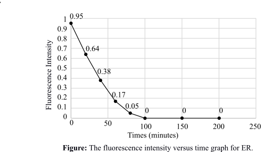
This graph depicts the intensity of fluorescence against time for the protein that is present in the ER. Initially, the fluorescence intensity is high, which means that the protein is entering the ER directly after synthesis. The graph plot is gradually decreasing, which is showing that the fluorescence in the ER is decreasing and reaching zero. This means that the fluorescent protein has moved out of the ER.
The graph of fluorescence intensity against time for the protein that is present in the Golgi apparatus is given below:
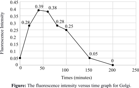
This graph depicts the intensity of fluorescence against time for the protein that is present in the Golgi apparatus. Initially, the fluorescence intensity is increasing, which means that the protein is entering the Golgi after a considerable time. The graph plot is gradually decreasing, which is showing that the fluorescence in the Golgi is decreasing and reaching zero. This means that the fluorescent protein is also moving out of the Golgi.
The graph of fluorescence intensity against time for the protein that is present in the cell membrane is given below:
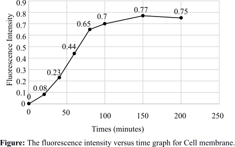
This graph depicts the intensity of fluorescence against time for the protein that is present in the cell membrane. The fluorescence intensity is increasing gradually and is maximized after some time, which means that the protein is embedded in the cell membrane.
The pathway that is followed by the protein is traced with the help of the graphs drawn. The protein enters the ER quickly after synthesis and then goes to the Golgi. From the Golgi, it moves to the cell membrane to form virus particles.
Therefore, the graphs are plotted and the pathway of the GFP fusion protein is traced with the help of the graphs. The pathway followed by the protein is from ER to Golgi and then to the cell membrane.
Want to see more full solutions like this?
Chapter 5 Solutions
Life: The Science of Biology


- Molecular Biology Please help with question. Thank you in advance. Discuss, compare and contrast the structure of promoters inprokaryotes and eukaryotes.arrow_forwardMolecular Biology Please help with question. Thank you You are studying the expression of the lac operon. You have isolated mutants as described below. In the absence of glucose, explain/describe what would happen, for each mutant, to the expression of the lac operon when you add lactose AND what would happen when the bacteria has used up all of the lactose (if the mutant is able to use lactose).1. Mutations in the lac repressor gene that would prevent the binding of lactose2. Mutations in the lac repressor gene that would prevent release of lactose once lactose hadbound3. Normally the lac repressor gene is located next to (a few hundred base pairs) and upstreamfrom the lac operon. Mutations in the lac repressor gene that move the lac repressor gene 100,000base pairs downstream.4. Mutations in the lac operator that would prevent binding of lac repressorarrow_forwardYou have returned to college to become a phylogeneticist. One of the first things you wish to do is determine how mammals, birds, and reptiles are related. Like any good scientist, you need to consider all available data objectively and without a preconceived “correct” answer. In pursuit of that, you should produce a phylogenetic tree based only on morphological features that show birds and mammals are more closely related. You will then produce a totally different tree, also using morphological features, that shows birds and reptiles are more closely related. Do not forget to include all three groups in both your trees. Based solely off the trees you produce, which relationship would you consider the more likely and why? Once you have answered that question, provide a brief summary of the “modern” understanding of the relationship between these three groups.arrow_forward
- true or false, the reason geckos can walk on walls is hydrogen bonding between their foot pads and the moisture on the wall.arrow_forwardBiology laboratory problem Please help. thank you You have 20 ul of DNA solution and 6X DNA loading buffer solution. You have to mix your DNA solution and DNA loading buffer before load DNA in an agarose gel. The concentration of the DNA loading buffer must be 1X in the DNA and DNA-loading buffer mixture after you mix them. For that, I will add _____ ul of 6X loading buffer to the 20 ul DNA solution.arrow_forwardBiology lab problem To make 20 ul of 5 mM MgCl2 solution using 50 mM MgCl2 stock solution and distilled water, I will mix ________ ul of 50 mM MgCl2 solution and ________ ul of distilled water. Please help . Thank youarrow_forward
- Biology Please help. Thank you. Biology laboratory question You need 50 ml of 1% (w/v) agarose gel. Agarose is a powder. How would you make it? You can ignore the volume of agarose powder. Don't forget the unit.TBE buffer is used to make an agarose gel, not distilled water. I will add _______ of agarose powder into 50 ml of distilled water (final 50 ml).arrow_forwardAn urgent care center experienced the average patient admissions shown in the Table below during the weeks from the first week of December through the second week of April. Week Average Daily Admissions 1-Dec 11 2-Dec 14 3-Dec 17 4-Dec 15 1-Jan 12 2-Jan 11 3-Jan 9 4-Jan 9 1-Feb 12 2-Feb 8 3-Feb 13 4-Feb 11 1-Mar 15 2-Mar 17 3-Mar 14 4-Mar 19 5-Mar 13 1-Apr 17 2-Apr 13 Forecast admissions for the periods from the first week of December through the second week of April. Compare the forecast admissions to the actual admissions; What do you conclude?arrow_forwardAnalyze the effectiveness of the a drug treatment program based on the needs of 18-65 year olds who are in need of treatment by critically describing 4 things in the program is doing effectively and 4 things the program needs some improvement.arrow_forward
- I have the first half finished... just need the bottom half.arrow_forward13. Practice Calculations: 3 colonies were suspended in the following dilution series and then a viable plate count and microscope count was performed. Calculate IDF's, TDF's and then calculate the CFU/mL in each tube by both methods. Finally calculate the cells in 1 colony by both methods. Show all of your calculations in the space provided on the following pages. 3 colonies 56 cells 10 μL 10 μL 100 μL 500 με m OS A B D 5.0 mL 990 με 990 με 900 με 500 μL EN 2 100 με 100 μL 118 colonies 12 coloniesarrow_forwardDescribe and give a specific example of how successionary stage is related to species diversity?arrow_forward


 Human Heredity: Principles and Issues (MindTap Co...BiologyISBN:9781305251052Author:Michael CummingsPublisher:Cengage Learning