Campbell Biology, Books a la Carte Plus Mastering Biology with eText -- Access Card Package (10th Edition)
10th Edition
ISBN: 9780133922851
Author: Jane B. Reece, Lisa A. Urry, Michael L. Cain, Steven A. Wasserman, Peter V. Minorsky, Robert B. Jackson
Publisher: PEARSON
expand_more
expand_more
format_list_bulleted
Concept explainers
Videos
Textbook Question
Chapter 47, Problem 8TYU
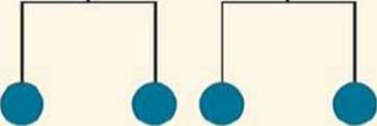
DRAW IT Each blue circle in the figure below represents a cell in a cell lineage. Draw two modified versions of the cell lineage so that each Version produces three cells. Use apoptosis in one of the versions, marking any dead cells with an X.

Expert Solution & Answer
Want to see the full answer?
Check out a sample textbook solution
Students have asked these similar questions
1. In vivo testing provides valuable insight into a drug’s kinetics. Assessing drug kinetics following multiple different routes of administration provides greater insight than just a single route of administration alone. The following data was collected in 250 g rats following bolus iv, oral (po), and intraperitoneal (ip) administration.Using this data and set of graphs, determine:
(a) k, C0, V, and AUC* for the bolus iv data (b) k, ka, B1, and AUC* for the po data (c) k, ka, B1, and AUC* for the ip data (d) relative bioavailability for po vs ip, Fpo/Fip (e) absolute po bioavailability, (f)Fpo absolute ip bioavailability, Fip
MAKE SURE ANSWERS HAVE UNITS if appropriate.
SHOW ALL WORK, including equation used, variables used and each step to your solution.
2. Drug quantification from plasma is commonly performed by using techniques such as HPLC or LC/MS. However, these methods do have limitations, and investigators may choose to use a radiolabeled analog of a drug instead. Radioligands are molecules that contain radioactive isotopes, commonly 3H or 14C. This technique allows investigators to quantify drug concentration from radiation measurements. The following measurements were made in 250 g rats following oral administration of 18.2 µCi of a 14C-labeled drug of interest: Time (min) Plasma Radiation Levels (µCi/L) 0 0.0 2 9.7 4 19.2 7 25.3 9 37.8 12 39.6 14 45.8 17 48.8 20 52.0 25 56.4 30 59.2 35 60.1 40 61.1 45 62.1 50 62.8 60 63.1 70 62.1 80 60.1 90 57.3 100 55.5 110 53.7 120 52.2 150 48.0 180 45.0 240 39.0
Note that a µCi is a measure of the amount of radioactivity and hence is a measure of the amount of drug present. Given that the oral bioavailability of this drug is known to be essentially 100%, estimate the following from this…
The current nutrition labelling regulation in Hong Kong requires food manufacturer to list E+7 information on the package of pre-packaged food products. Do you think that more nutrients, such as calcium and cholesterol, shall be included?
Chapter 47 Solutions
Campbell Biology, Books a la Carte Plus Mastering Biology with eText -- Access Card Package (10th Edition)
Ch. 47.1 - How does the fertilization envelope form in sea...Ch. 47.1 - Prob. 2CCCh. 47.1 - MAKE CONNECTIONS Review Figure 12.16 on cell...Ch. 47.2 - In the frog embryo, convergent extension elongates...Ch. 47.2 - WHAT IF? Predict what would happen if Ca2+ was...Ch. 47.2 - MAKE CONNECTIONS Unlike some other types of birth...Ch. 47.3 - Prob. 1CCCh. 47.3 - Prob. 2CCCh. 47.3 - Prob. 3CCCh. 47.3 - Prob. 4CC
Ch. 47 - What cell-surface event would likely fail if a...Ch. 47 - Prob. 47.2CRCh. 47 - Suppose you found two classes of mouse mutations,...Ch. 47 - Prob. 1TYUCh. 47 - Which of the following is common to the...Ch. 47 - The archenteron develops into a. the mesoderm. b....Ch. 47 - What structural adaptation in chickens allows them...Ch. 47 - Prob. 5TYUCh. 47 - In humans, identical twins are possible because a....Ch. 47 - Cells transplanted from the neural tube of a frog...Ch. 47 - DRAW IT Each blue circle in the figure below...Ch. 47 - EVOLUTION CONNECTION Evolution in insects and...Ch. 47 - Prob. 10TYUCh. 47 - Prob. 11TYUCh. 47 - Prob. 12TYUCh. 47 - SYNTHESIZE YOUR KNOWLEDGE Occasionally, two-headed...
Knowledge Booster


Learn more about
Need a deep-dive on the concept behind this application? Look no further. Learn more about this topic, biology and related others by exploring similar questions and additional content below.Similar questions
- View History Bookmarks Window Help Quarter cements ents ons (17) YouTube Which amino acids would you expect to find marked on the alpha helix? canvas.ucsc.edu ucsc Complaint and Grievance Process - Academic Personnel pach orations | | | | | | | | | | | | | | | | 000000 000000000 | | | | | | | | | | | | | | | | | | | | | | 00000000 scope vious De 48 12.415 KATPM FEB 3 F1 F2 80 F3 a F4 F5 2 # 3 $ 85 % tv N A の Mon Feb 3 10:24 PM Lipid bilayer Submit Assignment Next > ZOOM < Å DII 8 བ བ F6 16 F7 F8 F9 F10 34 F11 F12 & * ( 6 7 8 9 0 + 11 WERTY U { 0 } P deletearrow_forwardDifferent species or organisms research for ecologyarrow_forwardWhat is the result of the following gram stain: positive ○ capsulated ○ acid-fast ○ negativearrow_forward
- What type of stain is the image below: capsule stain endospore stain gram stain negative stain ASM MicrobeLibrary.org Keplingerarrow_forwardWhat is the result of the acid-fast stain below: Stock Images by Getty Images by Getty Images by Getty Images by Getty Image Getty Images St Soy Getty Images by Getty Images by Getty Images Joy Getty encapsulated O endosporulating negative ○ positivearrow_forwardYou have a stock vial of diligence 75mg in 3ml and need to draw up a dose of 50mg for your patient.how many mls should you draw up to give this dosearrow_forward
- You are recquired to administer 150mg hydrocortisone intravenously,how many mls should you give?(stock =hydrocortisone 100mg in 2mls)arrow_forwardIf someone was working with a 50 MBq F-18 source, what would be the internal and external dose consequences?arrow_forwardWe will be starting a group project next week where you and your group will research and ultimately present on a current research article related to the biology of a pathogen that infects humans. The article could be about the pathogen itself, the disease process related to the pathogen, the immune response to the pathogen, vaccines or treatments that affect the pathogen, or other biology-related study about the pathogen. I recommend that you choose a pathogen that is currently interesting to researchers, so that you will be able to find plenty of articles about it. Avoid choosing a historical disease that no longer circulates. List 3 possible pathogens or diseases that you might want to do for your group project.arrow_forward
- not use ai pleasearrow_forwardDNK dagi nukleotidlar va undan sintezlangan oqsildagi peptid boglar farqi 901 taga teng bo'lib undagi A jami H boglardan 6,5 marta kam bo'lsa DNK dagi jami H bog‘lar sonini topingarrow_forwardOne of the ways for a cell to generate ATP is through the oxidative phosphorylation. In oxidative phosphorylation 3 ATP are produced from every one NADH molecule. In respiration, every glucose molecule produces 10 NADH molecules. If a cell is growing on 5 glucose molecules, how much ATP can be produced using oxidative phosphorylation/aerobic respiration?arrow_forward
arrow_back_ios
SEE MORE QUESTIONS
arrow_forward_ios
Recommended textbooks for you
 Biology 2eBiologyISBN:9781947172517Author:Matthew Douglas, Jung Choi, Mary Ann ClarkPublisher:OpenStax
 Biology (MindTap Course List)BiologyISBN:9781337392938Author:Eldra Solomon, Charles Martin, Diana W. Martin, Linda R. BergPublisher:Cengage Learning
 Human Biology (MindTap Course List)BiologyISBN:9781305112100Author:Cecie Starr, Beverly McMillanPublisher:Cengage Learning
 Biology Today and Tomorrow without Physiology (Mi...BiologyISBN:9781305117396Author:Cecie Starr, Christine Evers, Lisa StarrPublisher:Cengage Learning
 Human Heredity: Principles and Issues (MindTap Co...BiologyISBN:9781305251052Author:Michael CummingsPublisher:Cengage Learning
Biology 2e
Biology
ISBN:9781947172517
Author:Matthew Douglas, Jung Choi, Mary Ann Clark
Publisher:OpenStax
Biology (MindTap Course List)
Biology
ISBN:9781337392938
Author:Eldra Solomon, Charles Martin, Diana W. Martin, Linda R. Berg
Publisher:Cengage Learning
Human Biology (MindTap Course List)
Biology
ISBN:9781305112100
Author:Cecie Starr, Beverly McMillan
Publisher:Cengage Learning
Biology Today and Tomorrow without Physiology (Mi...
Biology
ISBN:9781305117396
Author:Cecie Starr, Christine Evers, Lisa Starr
Publisher:Cengage Learning
Human Heredity: Principles and Issues (MindTap Co...
Biology
ISBN:9781305251052
Author:Michael Cummings
Publisher:Cengage Learning
The Cell Cycle and its Regulation; Author: Professor Dave Explains;https://www.youtube.com/watch?v=eqJqhA8HSJ0;License: Standard YouTube License, CC-BY
Cell Division - Mitosis and Meiosis - GCSE Biology (9-1); Author: Mr Exham Biology;https://www.youtube.com/watch?v=w7vp_uRA8kw;License: Standard YouTube License, CC-BY