Campbell Biology: Concepts & Connections (9th Edition)
9th Edition
ISBN: 9780134296012
Author: Martha R. Taylor, Eric J. Simon, Jean L. Dickey, Kelly A. Hogan, Jane B. Reece
Publisher: PEARSON
expand_more
expand_more
format_list_bulleted
Concept explainers
Textbook Question
Chapter 4, Problem 1CC
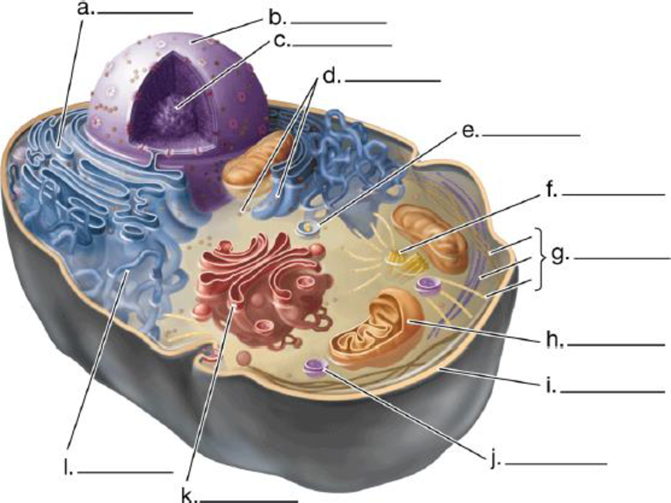
Label the structures in this diagram of an animal cell. Review the functions of each of these organelles.

Expert Solution & Answer
Summary Introduction
To review: The structures and functions of each of the organelles in the given animal cell.
Introduction:
A cell is the fundamental structural and functional unit of living organisms. It is bounded by a cell membrane. Cells contain different organelles. Each organelle has its own specific features and functions. Each cellular organelle possesses different structural peculiarities.
Explanation of Solution
Pictorial representation:
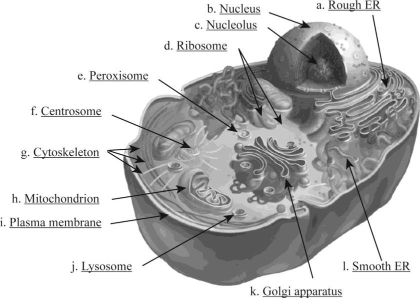
Fig. 1 shows various structural organelles of an animal cell.

Fig. 1: Structure of an animal cell
The functions of various organelles are very specific.
- a. Rough ER: It is a network of membranous sacs and tubes; its external surface is attached with the ribosome. It helps in protein processing and secretion. Also, the addition of carbohydrate with protein occurs to produce glycoprotein, which helps in the production of a new membrane within the cell.
- b. Nucleus: It is a membrane-bound organelle covered by the nuclear envelope. The nucleus contains the nucleolus and chromatin material. The chromatin material consists of DNA and proteins.
- c. Nucleolus: It is a core region in the nucleus where components of the ribosome assemble.
- d. Ribosome: It is a non-membranous structure found in the cytoplasm of the cell. It is the core organelle for protein synthesis. It can be found free or attached with the rough endoplasmic reticulum.
- e. Peroxisome: It is a specialized single membrane–bound vesicle found in the cytoplasm of the cell. It produces hydrogen peroxide as a by-product and converts it to water.
- f. Centrosome: The formation of microtubules initiates at the centrosome. It contains a pair of centrioles.
- g. Cytoskeleton: It consists of a network of fibrous substances within the cytoplasm of a cell. The cytoskeleton consists of three main fibers called microtubules, microfilaments, and intermediate filaments. It helps in providing rigidity and gives structural support to the cell.
- h. Mitochondrion: It is double membrane–bound organelle. It is important for cellular respiration and energy generation in the form of ATP. The two phospholipid bilayer membranes enclosed with the mitochondria play a great role in functioning of mitochondria. It is also called as the “power house” of the cell.
- i. Plasma membrane: It is the protective compartment of the cell. It is composed of a lipid bilayer.
- j. Lysosome: It is a single membrane–bound organelle. It is the storage unit of hydrolytic enzymes. Breakdown of indigested substances takes place in lysosomes, and cell organelles are recycled here.
- k. Golgi apparatus: It is a single membrane–bound organelle stack of the flattened membranous sac. It is the active unit of modification, sorting, and secretion of cell products.
- l. Smooth ER: It is a network of membranous sacs and tubes but not attached with the ribosome. It helps in detoxification and lipid synthesis. It helps in folding of protein and transferring of the synthesized protein.
Want to see more full solutions like this?
Subscribe now to access step-by-step solutions to millions of textbook problems written by subject matter experts!
Students have asked these similar questions
The following table is from Kumar et. al. Highly Selective Dopamine D3 Receptor (DR) Antagonists and Partial Agonists Based on Eticlopride and the D3R Crystal Structure: New Leads for Opioid Dependence Treatment. J. Med Chem 2016.
The following figure is from Caterina et al. The capsaicin receptor: a heat activated ion channel in the
pain pathway. Nature, 1997. Black boxes indicate capsaicin, white circles indicate resinferatoxin.
You are a chef in a fancy new science-themed restaurant. You have a recipe that calls for 1 teaspoon of resinferatoxin, but you feel uncomfortable serving foods with "toxins" in them. How much capsaicin could you substitute instead?
What protein is necessary for packaging acetylcholine into synaptic vesicles?
Chapter 4 Solutions
Campbell Biology: Concepts & Connections (9th Edition)
Ch. 4 - Label the structures in this diagram of an animal...Ch. 4 - Prob. 2TYKCh. 4 - Prob. 3TYKCh. 4 - Which of the following clues would tell you...Ch. 4 - Prob. 5TYKCh. 4 - What four cellular components are shared by...Ch. 4 - Describe two different ways in which cilia can...Ch. 4 - In which cell would you find the most lysosomes?...Ch. 4 - Prob. 9TYKCh. 4 - Prob. 10TYK
Ch. 4 - In which cell would you find the most...Ch. 4 - In what ways do the internal membranes of a...Ch. 4 - Is this statement true or false? Animal cells have...Ch. 4 - Describe the structure of the plasma membrane of...Ch. 4 - Prob. 15TYKCh. 4 - Describe the pathway of the protein hormone...Ch. 4 - How might the phrase ingested but not digested be...Ch. 4 - Cilia are found on cells in almost every organ of...Ch. 4 - SCIENTIFIC THINKING Microtubules often produce...
Knowledge Booster
Learn more about
Need a deep-dive on the concept behind this application? Look no further. Learn more about this topic, biology and related others by exploring similar questions and additional content below.Similar questions
- 1. Match each vocabulary term to its best descriptor A. affinity B. efficacy C. inert D. mimic E. how drugs move through body F. how drugs bind Kd Bmax Agonist Antagonist Pharmacokinetics Pharmacodynamicsarrow_forward50 mg dose of a drug is given orally to a patient. The bioavailability of the drug is 0.2. What is the volume of distribution of the drug if the plasma concentration is 1 mg/L? Be sure to provide units.arrow_forwardDetermine Kd and Bmax from the following Scatchard plot. Make sure to include units.arrow_forward
- Choose a catecholamine neurotransmitter and describe/draw the components of the synapse important for its signaling including synthesis, packaging into vesicles, receptors, transporters/degradative enzymes. Describe 2 drugs that can act on this system.arrow_forwardThe following figure is from Caterina et al. The capsaicin receptor: a heat activated ion channel in the pain pathway. Nature, 1997. Black boxes indicate capsaicin, white circles indicate resinferatoxin. a) Which has a higher potency? b) Which is has a higher efficacy? c) What is the approximate Kd of capsaicin in uM? (you can round to the nearest power of 10)arrow_forwardWhat is the rate-limiting-step for serotonin synthesis?arrow_forward
arrow_back_ios
SEE MORE QUESTIONS
arrow_forward_ios
Recommended textbooks for you
 Human Physiology: From Cells to Systems (MindTap ...BiologyISBN:9781285866932Author:Lauralee SherwoodPublisher:Cengage Learning
 Biology Today and Tomorrow without Physiology (Mi...BiologyISBN:9781305117396Author:Cecie Starr, Christine Evers, Lisa StarrPublisher:Cengage Learning


Human Physiology: From Cells to Systems (MindTap ...
Biology
ISBN:9781285866932
Author:Lauralee Sherwood
Publisher:Cengage Learning


Biology Today and Tomorrow without Physiology (Mi...
Biology
ISBN:9781305117396
Author:Cecie Starr, Christine Evers, Lisa Starr
Publisher:Cengage Learning
Dissection Basics | Types and Tools; Author: BlueLink: University of Michigan Anatomy;https://www.youtube.com/watch?v=-_B17pTmzto;License: Standard youtube license