
Videos
To label: The urinary structures given in the figure.
Introduction: The urinary system is the drainage system of the body for removing wastes in the form of urine. For this process, the parts of the urinary system should work together in coordination. The mammalian urinary system includes kidneys, ureters, urinary bladder, and urethra.
Explanation of Solution
Pictorial representation: The organs of the female urinary system are given in the Fig.1.
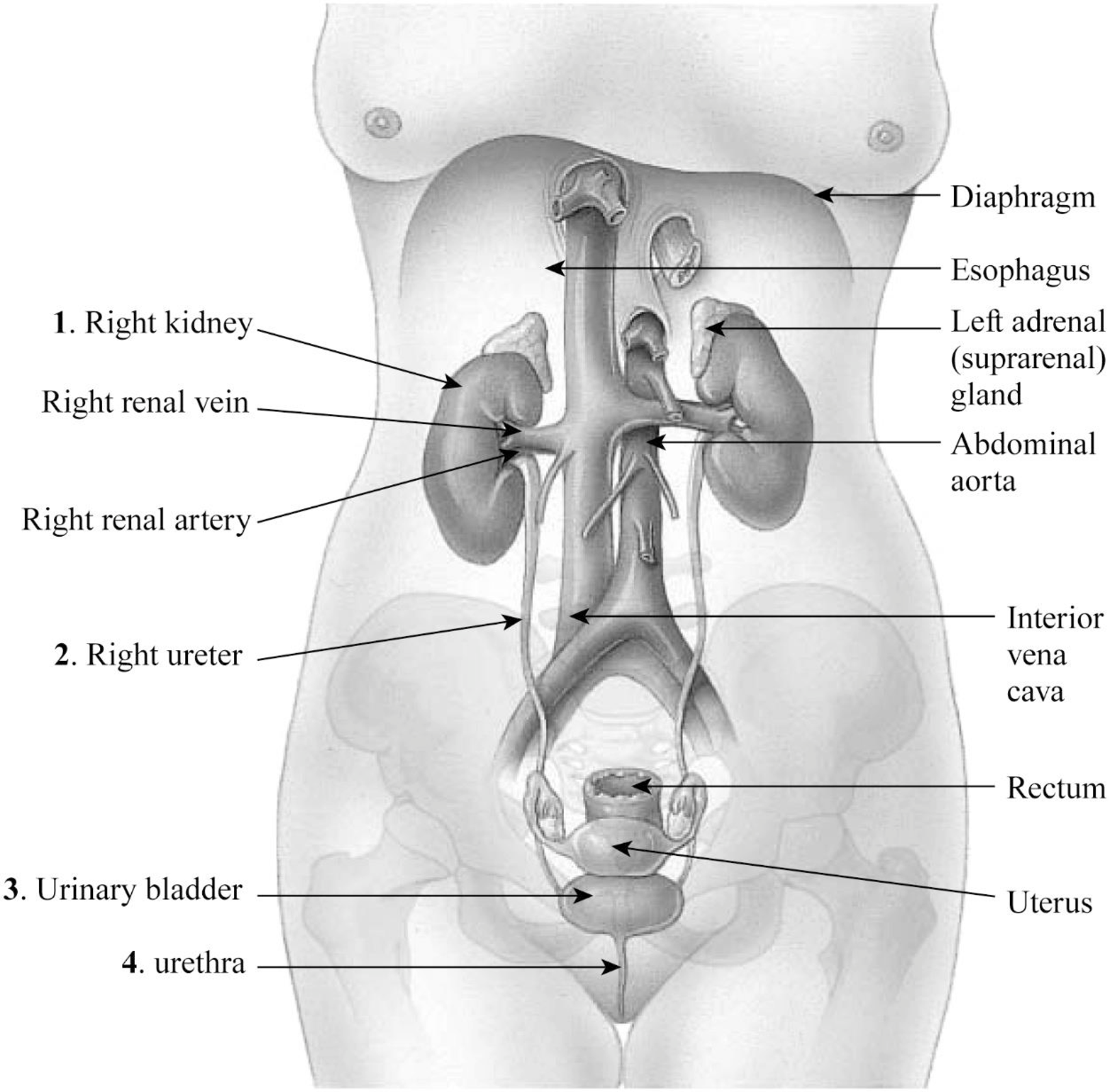
Fig.1: The organs of the female urinary system.
1. Right kidney: The kidney is an important organ of the excretory system. The right kidney is slightly longer than the left kidney.
2. Right ureter: The right ureter is located on the right side of the abdomen and is a long fibromuscular tube that transports urine from the right kidney to the urinary bladder. Urine makes the renal pelvis to contract in rhythm to drive urine through the ureters into the urinary bladder.
3. Urinary bladder: The wall of the urinary bladder has the detrusor muscle; it allows the storage of urine and contraction during urination and release of urine.
4. Urethra: Urethra is a tube that carries urine to the outside of the body. The bladder is joined to the urethra through the bladder neck that consists of internal sphincter muscles.
Pictorial representation: The location and coverings of the kidneys are given in Fig.2.
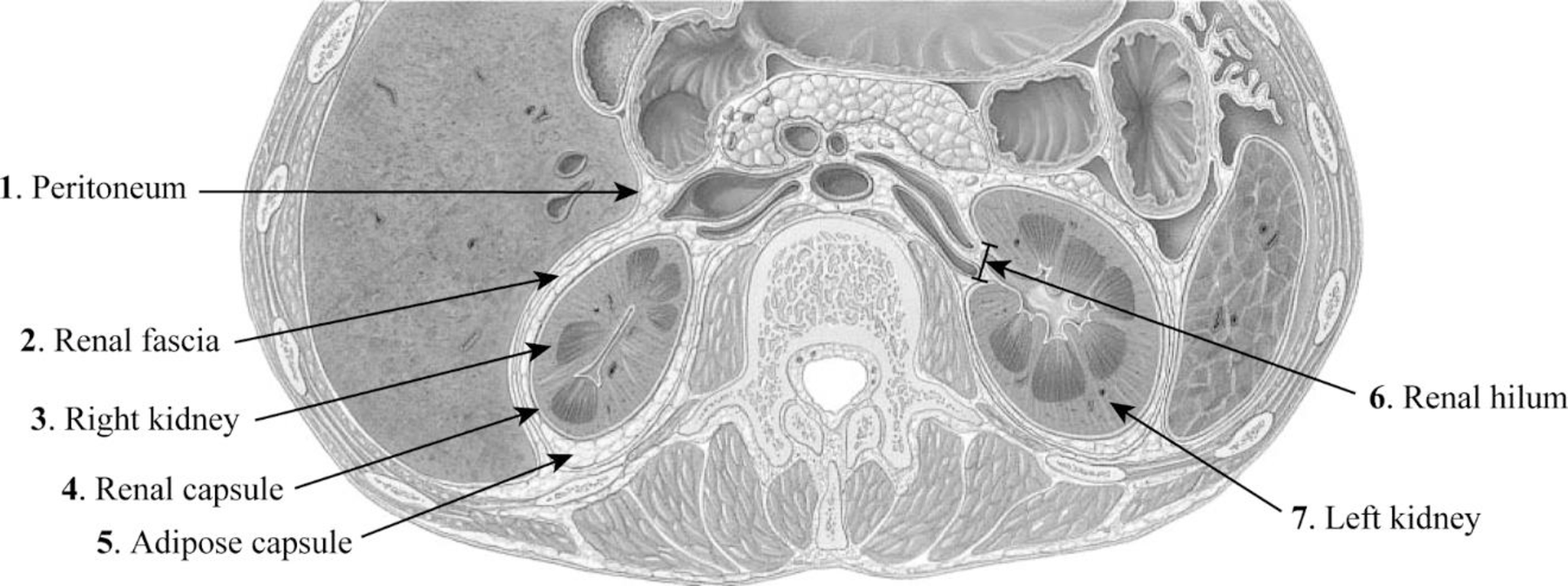
Fig.2: The location and coverings of the kidneys.
1. Peritoneum: The peritoneum is a sheet-like membrane that forms two layers and a space called the peritoneal cavity.
2. Renal fascia: The renal fascia is the layer of irregular connective tissue. This layer covers the adipose capsule and encapsulates the kidney. It passes anteriorly to the kidney, abdominal aorta, and renal vessels and posteriorly connects to the psoas fascia.
3. Right kidney: The right kidney is located on the right side of the abdomen and is connected to the right ureters through which urine reaches the urinary bladder.
4. Renal capsule: The fibrous capsule of the kidney is also known as the renal capsule. It is directly adhered to the external surface of the kidney.
5. Adipose capsule: The adipose capsule is the structure of the kidney that is located between the renal fascia and renal capsule. It is the perirenal fat that covers the renal capsule. This layer provides protection to the kidney from damage.
6. Renal hilum: The renal hilum is a depression in the surface of the kidney where blood vessels and nerves enter and exit.
7. Left kidney: The left kidney is located on the left side of the abdomen and is slightly shorter than the right kidney.
Pictorial representation: The internal structure of the kidney is given in Fig.3.
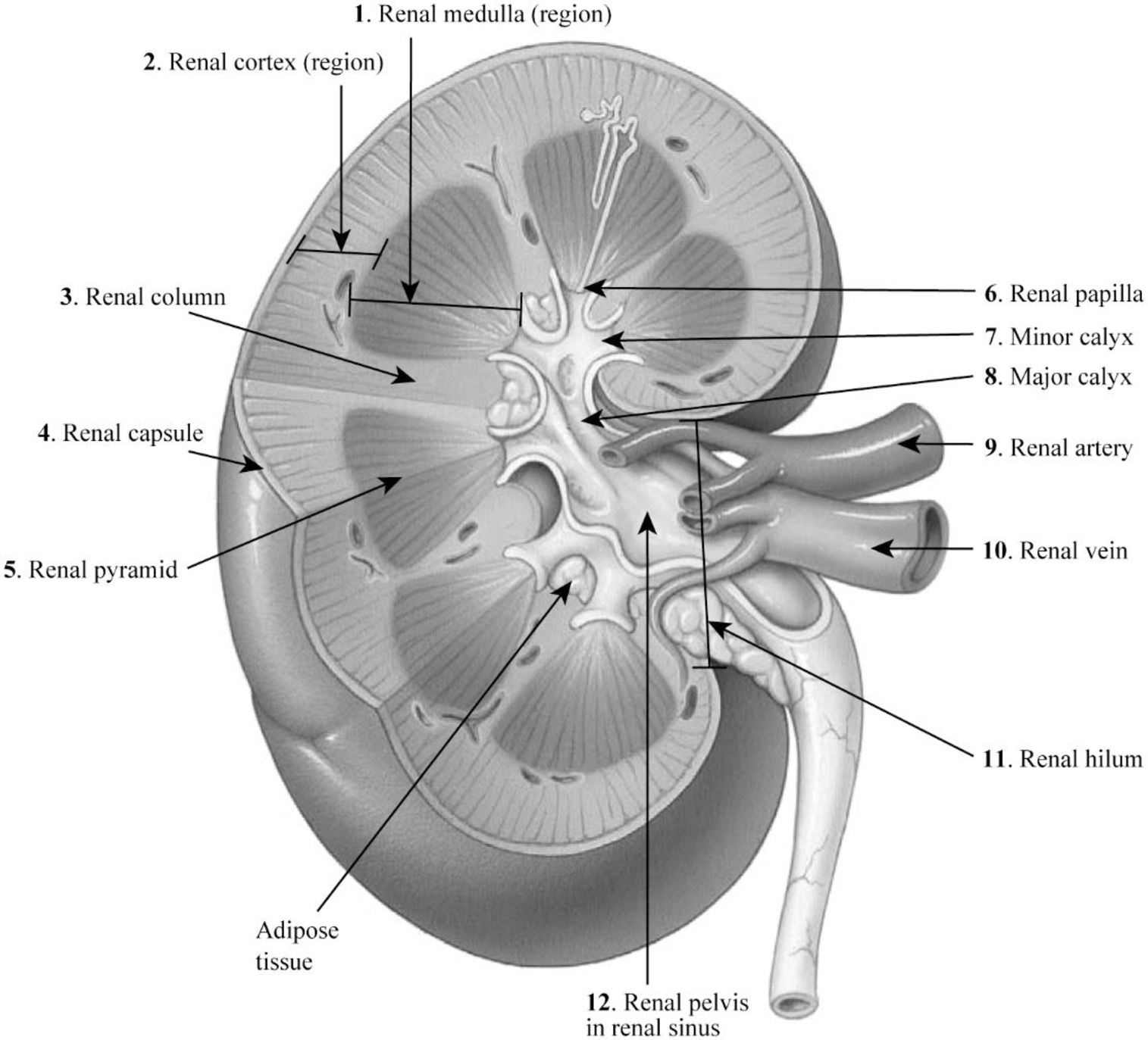
Fig.3: The internal structure of the kidney.
1. Renal medulla: The renal medulla is the deep region to the cortex of the kidney that contains 8–12 renal pyramids consisting of collecting ducts.
2. Renal cortex: The renal cortex is the outer portion of the kidney. It is a smooth outer zone present between the renal capsule and the renal medulla.
3. Renal column: The medullary renal cortex extensions present in between renal pyramids are called the renal column. This allows the renal cortex to be better attached, and each column consists of blood vessel and urinary tube lines.
4. Renal capsule: The renal capsule is composed of dense irregular connective tissue and maintains the kidney’s shape, protects from trauma, and prevents infectious pathogens from penetrating the kidney.
5. Renal pyramid: The renal pyramid is a cone-shaped tissue that consists of various tubules that transport located within the renal medulla. At the renal papilla, the renal pyramids drain urine into the minor calyx in the kidney.
6. Renal papilla: The renal papilla is the peak of the renal pyramid. At the renal papilla, the renal pyramids drain urine into the minor calyx in the kidney.
7. Minor calyx: The renal pelvis connects all the renal tubules and forms a cup-like region called calyces. The minor calyces surround the peak of the renal pyramids.
8. Major calyx: Two to three minor calyces unite to form the major calyx. Urine enters the papillary duct that is located within the renal papilla and then passes through the spaces (minor calyx, major calyx, and renal pelvis) within the renal sinus of the kidney.
9. Renal artery: The renal arteries carry a very large amount of blood from the heart to the kidneys for the process of filtration.
10. Renal vein: A large amount of blood enters the kidneys at the hilum through the renal artery and leaves the kidneys through the renal vein.
11. Renal hilum: A large amount of blood enters the kidneys at the hilum through the renal artery and leaves the kidneys through the renal vein.
12. Renal pelvis in renal sinus: The renal pelvis in the renal sinus connects all the renal tubules and forms a cup-like region called calyces. The minor calyces surround the peak of the renal pyramids. At the renal papilla, the renal pyramids drain urine into the minor calyx in the kidney.
Want to see more full solutions like this?
Chapter 36 Solutions
EBK LABORATORY MANUAL FOR ANATOMY AND P


- 9. Aerobic respiration of one lipid molecule. The lipid is composed of one glycerol molecule connected to two fatty acid tails. One fatty acid is 12 carbons long and the other fatty acid is 18 carbons long in the figure below. Use the information below to determine how much ATP will be produced from the glycerol part of the lipid. Then, in part B, determine how much ATP is produced from the 2 fatty acids of the lipid. Finally put the NADH and ATP yields together from the glycerol and fatty acids (part A and B) to determine your total number of ATP produced per lipid. Assume no other carbon source is available. 18 carbons fatty acids 12 carbons 9 glycerol A. Glycerol is broken down to glyceraldehyde 3-phosphate, a glycolysis intermediate via the following pathway shown in the figure below. Notice this process costs one ATP but generates one FADH2. Continue generating ATP with glyceraldehyde-3-phosphate using the standard pathway and aerobic respiration. glycerol glycerol-3- phosphate…arrow_forwardNormal dive (for diving humans) normal breathing dive normal breathing Oz level CO2 level urgent need to breathe Oz blackout zone high CO2 triggers breathing 6. This diagram shows rates of oxygen depletion and carbon dioxide accumulation in the blood in relation to the levels needed to maintain consciousness and trigger the urgent need to breathe in diving humans. How might the location and slope of the O₂ line differ for diving marine mammals such as whales and dolphins? • How might the location and slope of the CO₂ line differ for diving marine mammals such as whales and dolphins? • • Draw in predicted lines for O2 and CO2, based on your reasoning above. How might the location of the Urgent Need to Breathe line and the O2 Blackout Zone line differ for diving marine mammals? What physiological mechanisms account for each of these differences, resulting in the ability of marine mammals to stay submerged for long periods of time?arrow_forwardforaging/diet type teeth tongue stomach intestines cecum Insectivory numerous, spiky, incisors procumbentExample: moleExample: shrew -- simple short mostly lacking Myrmecophagy absent or reduced in numbers, peg-likeExample: tamandua anteater extremely long simple, often roughened short small or lacking Terrestrial carnivory sharp incisors; long, conical canines; often carnassial cheek teeth; may have crushing molarsExample: dog -- simple short small Aquatic carnivory homodont, spiky, numerousExample: common dolphin -- simple or multichambered (cetaceans only) variable small or absent Sanguinivory very sharp upper incisors; reduced cheek teethExample: vampire bat grooved tubular, highly extensible long small or lacking Herbivory (except nectivores) incisors robust or absent; canines reduced or absent; diastema; cheek teeth enlarged with complex occlusal surfacesExample: beaver -- simple (hindgut fermenters) or multichambered (ruminants) long large Filter feeding none…arrow_forward
- 3. Shown below is the dental formula and digestive tract anatomy of three mammalian species (A, B, and C). What kind of diet would you expect each species to have? Support your answers with what you can infer from the dental formula and what you can see in the diagram. Broadly speaking, what accounts for the differences? Species A 3/3, 1/1, 4/4, 3/3 པར『ན་ cm 30 Species B 4/3, 1/1, 2/2, 4/4 cm 10 Species C 0/4, 0/0,3/3, 3/3 020arrow_forward3. Shown below is the dental formula and digestive tract anatomy of three mammalian species (A, B, and C). What kind of diet would you expect each species to have? Support your answers with what you can infer from the dental formula and what you can see in the diagram. Broadly speaking, what accounts for the differences? Species A 3/3, 1/1, 4/4, 3/3 cm 30 Species B 0/4, 0/0, 3/3, 3/3 cm 10 Species C 4/3, 1/1, 2/2, 4/4 E 0 cm 20 AILarrow_forwardNormal dive (for diving humans) normal breathing dive normal breathing Oz level CO₂ level urgent need to breathe Oz blackout zone high CO₂ triggers breathing 6. This diagram shows rates of oxygen depletion and carbon dioxide accumulation in the blood in relation to the levels needed to maintain consciousness and trigger the urgent need to breathe in diving humans. • How might the location and slope of the O2 line differ for diving marine mammals such as whales and dolphins? • How might the location and slope of the CO2 line differ for diving marine mammals such as whales and dolphins? • • Draw in predicted lines for O2 and CO2, based on your reasoning above. How might the location of the Urgent Need to Breathe line and the O2 Blackout Zone line differ for diving marine mammals? What physiological mechanisms account for each of these differences, resulting in the ability of marine mammals to stay submerged for long periods of time?arrow_forward
- How much ATP will be produced during the following metabolic scenario: Aerobic respiration of a 5mM lipid solution that is made up of one glycerol and an 8-carbon fatty acid and 12-carbon fatty acid. Recall that when glycerol breaks down to Glyceraldehyde-3-phosphate it costs one ATP but your get an extra FADH2. Every two carbons of a fatty acid break down to one acetyl-CoA. Units cannot be entered in this style of question but the units of your answer should be in mM of ATP.arrow_forwardIf a bacterium using aerobic respiration was to degrade one small protein molecule into 8 molecules of pyruvic acid, how many ATP would that cell make? Assume there is no other carbon source. Units cannot be entered in this style of question but the units of your answer should be in molecules of ATP.arrow_forwardIf a bacterium using aerobic respiration was to degrade a 30 mM solution of citric acid, how many ATP would that cell make? Assume no other carbon source is available. Units cannot be entered in this style of question but the units of your answer should be in mM of ATP.arrow_forward
- How much ATP will be produced during the following metabolic scenario: Aerobic respiration of a 5mM lipid solution that is made up of one glycerol and an 8-carbon fatty acid and 12-carbon fatty acid. Recall that when glycerol breaks down to Glyceraldehyde-3-phosphate it costs one ATP but your get an extra FADH2. Every two carbons of a fatty acid break down to one acetyl-CoA. (pathways will be provided on the exam) Units cannot be entered in this style of question but the units of your answer should be in mM of ATP.arrow_forwardWhen beta-lactamase was isolated from Staphylcoccus aureus and treated with a phosphorylating agent, only the active site, serine was phosphorylated. Additionally, the serine was found to constitute 0.35% (by weight) of this beta-lactamase enzyme. Using this, calculate the molecular weight of this enzyme and estimate the number of amino acids present in the polypeptide.arrow_forwardBased on your results from the Mannitol Salt Agar (MSA) media, which of your bacteria were mannitol fermenters and which were not mannitol fermenters?arrow_forward
 Human Anatomy & Physiology (11th Edition)BiologyISBN:9780134580999Author:Elaine N. Marieb, Katja N. HoehnPublisher:PEARSON
 Biology 2eBiologyISBN:9781947172517Author:Matthew Douglas, Jung Choi, Mary Ann ClarkPublisher:OpenStax
 Anatomy & PhysiologyBiologyISBN:9781259398629Author:McKinley, Michael P., O'loughlin, Valerie Dean, Bidle, Theresa StouterPublisher:Mcgraw Hill Education,
 Molecular Biology of the Cell (Sixth Edition)BiologyISBN:9780815344322Author:Bruce Alberts, Alexander D. Johnson, Julian Lewis, David Morgan, Martin Raff, Keith Roberts, Peter WalterPublisher:W. W. Norton & Company
 Laboratory Manual For Human Anatomy & PhysiologyBiologyISBN:9781260159363Author:Martin, Terry R., Prentice-craver, CynthiaPublisher:McGraw-Hill Publishing Co.
 Inquiry Into Life (16th Edition)BiologyISBN:9781260231700Author:Sylvia S. Mader, Michael WindelspechtPublisher:McGraw Hill Education