Videos
To label: The urinary structures given in the figure.
Introduction: The urinary system is the drainage system of the body for removing wastes in the form of urine. For this process, the parts of the urinary system should work together in coordination. The mammalian urinary system includes kidneys, ureters, urinary bladder, and urethra.
Explanation of Solution
Pictorial representation: The organs of the female urinary system are given in the Fig.1.
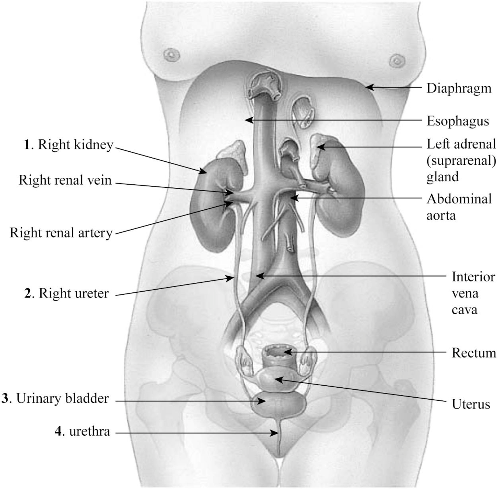
Fig.1: The organs of the female urinary system.
1. Right kidney: The kidney is an important organ of the excretory system. The right kidney is slightly longer than the left kidney.
2. Right ureter: The right ureter is located on the right side of the abdomen and is a long fibromuscular tube that transports urine from the right kidney to the urinary bladder. Urine makes the renal pelvis to contract in rhythm to drive urine through the ureters into the urinary bladder.
3. Urinary bladder: The wall of the urinary bladder has the detrusor muscle; it allows the storage of urine and contraction during urination and release of urine.
4. Urethra: Urethra is a tube that carries urine to the outside of the body. The bladder is joined to the urethra through the bladder neck that consists of internal sphincter muscles.
Pictorial representation: The location and coverings of the kidneys are given in Fig.2.
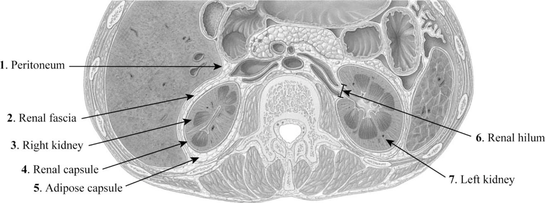
Fig.2: The location and coverings of the kidneys.
1. Peritoneum: The peritoneum is a sheet-like membrane that forms two layers and a space called the peritoneal cavity.
2. Renal fascia: The renal fascia is the layer of irregular connective tissue. This layer covers the adipose capsule and encapsulates the kidney. It passes anteriorly to the kidney, abdominal aorta, and renal vessels and posteriorly connects to the psoas fascia.
3. Right kidney: The right kidney is located on the right side of the abdomen and is connected to the right ureters through which urine reaches the urinary bladder.
4. Renal capsule: The fibrous capsule of the kidney is also known as the renal capsule. It is directly adhered to the external surface of the kidney.
5. Adipose capsule: The adipose capsule is the structure of the kidney that is located between the renal fascia and renal capsule. It is the perirenal fat that covers the renal capsule. This layer provides protection to the kidney from damage.
6. Renal hilum: The renal hilum is a depression in the surface of the kidney where blood vessels and nerves enter and exit.
7. Left kidney: The left kidney is located on the left side of the abdomen and is slightly shorter than the right kidney.
Pictorial representation: The internal structure of the kidney is given in Fig.3.
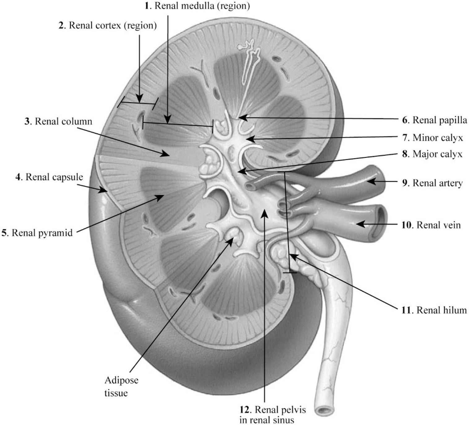
Fig.3: The internal structure of the kidney.
1. Renal medulla: The renal medulla is the deep region to the cortex of the kidney that contains 8–12 renal pyramids consisting of collecting ducts.
2. Renal cortex: The renal cortex is the outer portion of the kidney. It is a smooth outer zone present between the renal capsule and the renal medulla.
3. Renal column: The medullary renal cortex extensions present in between renal pyramids are called the renal column. This allows the renal cortex to be better attached, and each column consists of blood vessel and urinary tube lines.
4. Renal capsule: The renal capsule is composed of dense irregular connective tissue and maintains the kidney’s shape, protects from trauma, and prevents infectious pathogens from penetrating the kidney.
5. Renal pyramid: The renal pyramid is a cone-shaped tissue that consists of various tubules that transport located within the renal medulla. At the renal papilla, the renal pyramids drain urine into the minor calyx in the kidney.
6. Renal papilla: The renal papilla is the peak of the renal pyramid. At the renal papilla, the renal pyramids drain urine into the minor calyx in the kidney.
7. Minor calyx: The renal pelvis connects all the renal tubules and forms a cup-like region called calyces. The minor calyces surround the peak of the renal pyramids.
8. Major calyx: Two to three minor calyces unite to form the major calyx. Urine enters the papillary duct that is located within the renal papilla and then passes through the spaces (minor calyx, major calyx, and renal pelvis) within the renal sinus of the kidney.
9. Renal artery: The renal arteries carry a very large amount of blood from the heart to the kidneys for the process of filtration.
10. Renal vein: A large amount of blood enters the kidneys at the hilum through the renal artery and leaves the kidneys through the renal vein.
11. Renal hilum: A large amount of blood enters the kidneys at the hilum through the renal artery and leaves the kidneys through the renal vein.
12. Renal pelvis in renal sinus: The renal pelvis in the renal sinus connects all the renal tubules and forms a cup-like region called calyces. The minor calyces surround the peak of the renal pyramids. At the renal papilla, the renal pyramids drain urine into the minor calyx in the kidney.
Want to see more full solutions like this?
Chapter 36 Solutions
Laboratory Manual for Anatomy and Physiology, 6e Loose-Leaf Print Companion


- Transcription and Translation 1. What is the main function of transcription and translation? (2 marks) 2. How is transcription different in eukaryotic and prokaryotic cells? (2 marks) 3. Explain the difference between pre-mRNA and post-transcript mRNA. (2 marks) 4. What is the function of the following: (4 marks) i. the cap ii. spliceosome iii. Poly A tail iv. termination sequence 5. What are advantages to the wobble feature of the genetic code? (2 marks) 6. Explain the difference between the: (3 marks) i. A site & P site ii. codon & anticodon iii. gene expression and gene regulation 7. Explain how the stop codon allows for termination. (1 mark) 8. In your own words, summarize the process of translation. (2 marks)arrow_forwardIn this activity you will research performance enhancers that affect the endocrine system or nervous system. You will submit a 1 page paper on one performance enhancer of your choice. Be sure to include: the specific reason for use the alleged results on improving performance how it works how it affect homeostasis and improves performance any side-effects of this substancearrow_forwardNeurons and Reflexes 1. Describe the function of the: a) dendrite b) axon c) cell body d) myelin sheath e) nodes of Ranvier f) Schwann cells g) motor neuron, interneuron and sensory neuron 2. List some simple reflexes. Explain why babies are born with simple reflexes. What are they and why are they necessary. 3. Explain why you only feel pain after a few seconds when you touch something very hot but you have already pulled your hand away. 4. What part of the brain receives sensory information? What part of the brain directs you to move your hand away? 5. In your own words describe how the axon fires.arrow_forward
- Mutations Here is your template DNA strand: CTT TTA TAG TAG ATA CCA CAA AGG 1. Write out the complementary mRNA that matches the DNA above. 2. Write the anticodons and the amino acid sequence. 3. Change the nucleotide in position #15 to C. 4. What type of mutation is this? 5. Repeat steps 1 & 2. 6. How has this change affected the amino acid sequence? 7. Now remove nucleotides 13 through 15. 8. Repeat steps 1 & 2. 9. What type of mutation is this? 0. Do all mutations result in a change in the amino acid sequence? 1. Are all mutations considered bad? 2. The above sequence codes for a genetic disorder called cystic fibrosis (CF). 3. When A is changed to G in position #15, the person does not have CF. When T is changed to C in position #14, the person has the disorder. How could this have originated?arrow_forwardhoose a scientist(s) and research their contribution to our derstanding of DNA structure or replication. Write a one page port and include: their research where they studied and the time period in which they worked their experiments and results the contribution to our understanding of DNA cientists Watson & Crickarrow_forwardhoose a scientist(s) and research their contribution to our derstanding of DNA structure or replication. Write a one page port and include: their research where they studied and the time period in which they worked their experiments and results the contribution to our understanding of DNA cientists Watson & Crickarrow_forward
- 7. Aerobic respiration of a protein that breaks down into 12 molecules of malic acid. Assume there is no other carbon source and no acetyl-CoA. NADH FADH2 OP ATP SLP ATP Total ATP Show your work using dimensional analysis here: 3arrow_forwardFor each of the following problems calculate the following: (Week 6-3 Video with 6-1 and 6-2) Consult the total catabolic pathways on the last page as a reference for the following questions. A. How much NADH and FADH2 is produced and fed into the electron transport chain (If any)? B. How much ATP is made from oxidative phosphorylation (OP), if any? Feed the NADH and FADH2 into the electron transport chain: 3ATP/NADH, 2ATP/FADH2 C. How much ATP is made by substrate level phosphorylation (SLP)? D. How much total ATP is made? Add the SLP and OP together. 1. Aerobic respiration using 0.5 mole of glucose? NADH FADH2 OP ATP SLP ATP Total ATP Show your work using dimensional analysis here:arrow_forwardAerobic respiration of one lipid molecule. The lipid is composed of one glycerol molecule connected to two fatty acid tails. One fatty acid is 12 carbons long and the other fatty acid is 18 carbons long in the figure below. Use the information below to determine how much ATP will be produced from the glycerol part of the lipid. Then, in part B, determine how much ATP is produced from the 2 fatty acids of the lipid. Finally put the NADH and ATP yields together from the glycerol and fatty acids (part A and B) to determine your total number of ATP produced per lipid. Assume no other carbon source is available. 18 carbons fatty acids 12 carbons glycerol . Glycerol is broken down to glyceraldehyde 3-phosphate, a glycolysis intermediate via the following pathway shown in the figure below. Notice this process costs one ATP but generates one FADH2. Continue generating ATP with glyceraldehyde-3-phosphate using the standard pathway and aerobic respiration. glycerol glycerol-3- phosphate…arrow_forward
- Don't copy the other answerarrow_forward4. Aerobic respiration of 5 mM acetate solution. Assume no other carbon source and that acetate is equivalent to acetyl-CoA. NADH FADH2 OP ATP SLP ATP Total ATP Show your work using dimensional analysis here: 5. Aerobic respiration of 2 mM alpha-ketoglutaric acid solution. Assume no other carbon source. NADH FADH2 OP ATP Show your work using dimensional analysis here: SLP ATP Total ATParrow_forwardBiology You’re going to analyze 5 ul of your PCR product(out of 50 ul) on the gel. How much of 6X DNAloading buffer (dye) are you going to mix with yourPCR product to make final 1X concentration ofloading buffer in the PCR product-loading buffermixture?arrow_forward
 Human Anatomy & Physiology (11th Edition)BiologyISBN:9780134580999Author:Elaine N. Marieb, Katja N. HoehnPublisher:PEARSON
 Biology 2eBiologyISBN:9781947172517Author:Matthew Douglas, Jung Choi, Mary Ann ClarkPublisher:OpenStax
 Anatomy & PhysiologyBiologyISBN:9781259398629Author:McKinley, Michael P., O'loughlin, Valerie Dean, Bidle, Theresa StouterPublisher:Mcgraw Hill Education,
 Molecular Biology of the Cell (Sixth Edition)BiologyISBN:9780815344322Author:Bruce Alberts, Alexander D. Johnson, Julian Lewis, David Morgan, Martin Raff, Keith Roberts, Peter WalterPublisher:W. W. Norton & Company
 Laboratory Manual For Human Anatomy & PhysiologyBiologyISBN:9781260159363Author:Martin, Terry R., Prentice-craver, CynthiaPublisher:McGraw-Hill Publishing Co.
 Inquiry Into Life (16th Edition)BiologyISBN:9781260231700Author:Sylvia S. Mader, Michael WindelspechtPublisher:McGraw Hill Education