To analyze:
Major structures of the ear in thegiven diagram.
Introduction:
Ear is the sensory organ responsible for hearing in an individual. Due to differences in the air pressure of the surrounding area or even moderate level vibrations causes tympanic membrane to vibrate and thereby the waves proceed towards the inner ear, cochlea and hence enabling an organism to hear. The normal hearing range is 20 − 20000Hz andhuman beings can hear at 0 dB (decibel) frequency..
Explanation of Solution
The major structures in the ear are labeled.
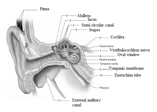
Ear comprises three bones which are known as ear ossicles. They are malleus, incus and stapes. These bones are interconnected to each other. Malleus is the first among the three ear ossicles. Pinna is the external part of outer ear. It is responsible for the collection of vibrations in the air which produce sound.Malleus is known as hammer bone because of its shape. It is attached to incus and when sound waves reach malleus, it transmits these waves from incus to the eardrum.Incus is the second bone among the ear ossicles. It is the connecting link between malleus and stapes.Incus is shaped like an anvil. Its one part which articulates with the malleus is known as short crus and the other part which is long and articulates with stapes is known as long crus. It receives the sound waves from the malleus and transmits it to the stapes for further processing.The inner ear comprises of a vestibular apparatus which comprises of the bony labyrinth part along with these semicircular canals or semicircular ducts and otolith organs. The inner ear has fluid filled labyrinth.
The sound waves or vibrations from the stapes reach cochlea. It is surrounded by membrane and the fluid present in its surrounding known as perilymph. This cochlea possesses hair cells or stereocilia which receives the sound waves and then further these sound waves produce nerve impulse which travels to the auditory cortex of the brain and thereby on being analyzed, sound can be heard.The vestibulocochlear nerve comprises of the cochlear nerve and the vestibular nerve. The name itself suggests that this nerve carries information about hearing as well as the balance of the body with regards to the posture. Thus, it performs a dual function.
Oval window connects the three ears ossicles present in the middle ear − malleus, incus and stapes to the cochlea of the ear. Stapes is the bone which gets connected to this oval window, but since the three bones are interconnected, therefore we say that ear ossicles are connected to the cochlea through this window.The tympanic membrane is composed of connective tissue. It receives vibrations from the external meatus and transmits them towards the ossicles. Also, it sets the demarcation between the outer ear and the middle ear.The tympanic membrane is composed of connective tissue. It receives vibrations from the external meatus and transmits them towards the ossicles. Also, it sets the demarcation between the outer ear and the middle ear.
Eustachian tube connects the middle ear to the nasopharynx thereby to the upper part of the throat. Eustachian tube has a major role in maintaining the balance of the body. It helps in equalizing air pressure between the middle ear and the external surrounding.External auditory canal comprises of hair and wax secreting sebaceous glands which help in trapping any dust particles present in the surrounding air which has to be passed to the middle ear. Thus, it helps in ensuring that purified air reaches the middle ear thereby preventing any sort of infection.
Want to see more full solutions like this?
Chapter 32 Solutions
Laboratory Manual for Holes Human Anatomy & Physiology Fetal Pig Version

- foraging/diet type teeth tongue stomach intestines cecum Insectivory numerous, spiky, incisors procumbentExample: moleExample: shrew -- simple short mostly lacking Myrmecophagy absent or reduced in numbers, peg-likeExample: tamandua anteater extremely long simple, often roughened short small or lacking Terrestrial carnivory sharp incisors; long, conical canines; often carnassial cheek teeth; may have crushing molarsExample: dog -- simple short small Aquatic carnivory homodont, spiky, numerousExample: common dolphin -- simple or multichambered (cetaceans only) variable small or absent Sanguinivory very sharp upper incisors; reduced cheek teethExample: vampire bat grooved tubular, highly extensible long small or lacking Herbivory (except nectivores) incisors robust or absent; canines reduced or absent; diastema; cheek teeth enlarged with complex occlusal surfacesExample: beaver -- simple (hindgut fermenters) or multichambered (ruminants) long large Filter feeding none…arrow_forward3. Shown below is the dental formula and digestive tract anatomy of three mammalian species (A, B, and C). What kind of diet would you expect each species to have? Support your answers with what you can infer from the dental formula and what you can see in the diagram. Broadly speaking, what accounts for the differences? Species A 3/3, 1/1, 4/4, 3/3 པར『ན་ cm 30 Species B 4/3, 1/1, 2/2, 4/4 cm 10 Species C 0/4, 0/0,3/3, 3/3 020arrow_forward3. Shown below is the dental formula and digestive tract anatomy of three mammalian species (A, B, and C). What kind of diet would you expect each species to have? Support your answers with what you can infer from the dental formula and what you can see in the diagram. Broadly speaking, what accounts for the differences? Species A 3/3, 1/1, 4/4, 3/3 cm 30 Species B 0/4, 0/0, 3/3, 3/3 cm 10 Species C 4/3, 1/1, 2/2, 4/4 E 0 cm 20 AILarrow_forward
- Normal dive (for diving humans) normal breathing dive normal breathing Oz level CO₂ level urgent need to breathe Oz blackout zone high CO₂ triggers breathing 6. This diagram shows rates of oxygen depletion and carbon dioxide accumulation in the blood in relation to the levels needed to maintain consciousness and trigger the urgent need to breathe in diving humans. • How might the location and slope of the O2 line differ for diving marine mammals such as whales and dolphins? • How might the location and slope of the CO2 line differ for diving marine mammals such as whales and dolphins? • • Draw in predicted lines for O2 and CO2, based on your reasoning above. How might the location of the Urgent Need to Breathe line and the O2 Blackout Zone line differ for diving marine mammals? What physiological mechanisms account for each of these differences, resulting in the ability of marine mammals to stay submerged for long periods of time?arrow_forwardHow much ATP will be produced during the following metabolic scenario: Aerobic respiration of a 5mM lipid solution that is made up of one glycerol and an 8-carbon fatty acid and 12-carbon fatty acid. Recall that when glycerol breaks down to Glyceraldehyde-3-phosphate it costs one ATP but your get an extra FADH2. Every two carbons of a fatty acid break down to one acetyl-CoA. Units cannot be entered in this style of question but the units of your answer should be in mM of ATP.arrow_forwardIf a bacterium using aerobic respiration was to degrade one small protein molecule into 8 molecules of pyruvic acid, how many ATP would that cell make? Assume there is no other carbon source. Units cannot be entered in this style of question but the units of your answer should be in molecules of ATP.arrow_forward
- If a bacterium using aerobic respiration was to degrade a 30 mM solution of citric acid, how many ATP would that cell make? Assume no other carbon source is available. Units cannot be entered in this style of question but the units of your answer should be in mM of ATP.arrow_forwardHow much ATP will be produced during the following metabolic scenario: Aerobic respiration of a 5mM lipid solution that is made up of one glycerol and an 8-carbon fatty acid and 12-carbon fatty acid. Recall that when glycerol breaks down to Glyceraldehyde-3-phosphate it costs one ATP but your get an extra FADH2. Every two carbons of a fatty acid break down to one acetyl-CoA. (pathways will be provided on the exam) Units cannot be entered in this style of question but the units of your answer should be in mM of ATP.arrow_forwardWhen beta-lactamase was isolated from Staphylcoccus aureus and treated with a phosphorylating agent, only the active site, serine was phosphorylated. Additionally, the serine was found to constitute 0.35% (by weight) of this beta-lactamase enzyme. Using this, calculate the molecular weight of this enzyme and estimate the number of amino acids present in the polypeptide.arrow_forward
- Based on your results from the Mannitol Salt Agar (MSA) media, which of your bacteria were mannitol fermenters and which were not mannitol fermenters?arrow_forwardhelp tutor pleasearrow_forwardQ8. A researcher wants to study the effectiveness of a pill intended to reduce stomach heartburn in pregnant women. The researcher chooses randomly 400 women to participate in this experiment for 9 months of their pregnancy period. They all need to have the same diet. The researcher designs two groups of 200 participants: One group take the real medication intended to reduce heartburn, while the other group take placebo medication. In this study what are: Independent variable: Dependent variable: Control variable: Experimental group: " Control group: If the participants do not know who is consuming the real pills and who is consuming the sugar pills. This study is It happens that 40% of the participants do not find the treatment helpful and drop out after 6 months. The researcher throws out the data from subjects that drop out. What type of bias is there in this study? If the company who makes the medication funds this research, what type of bias might exist in this research work?arrow_forward
 Human Anatomy & Physiology (11th Edition)BiologyISBN:9780134580999Author:Elaine N. Marieb, Katja N. HoehnPublisher:PEARSON
 Biology 2eBiologyISBN:9781947172517Author:Matthew Douglas, Jung Choi, Mary Ann ClarkPublisher:OpenStax
 Anatomy & PhysiologyBiologyISBN:9781259398629Author:McKinley, Michael P., O'loughlin, Valerie Dean, Bidle, Theresa StouterPublisher:Mcgraw Hill Education,
 Molecular Biology of the Cell (Sixth Edition)BiologyISBN:9780815344322Author:Bruce Alberts, Alexander D. Johnson, Julian Lewis, David Morgan, Martin Raff, Keith Roberts, Peter WalterPublisher:W. W. Norton & Company
 Laboratory Manual For Human Anatomy & PhysiologyBiologyISBN:9781260159363Author:Martin, Terry R., Prentice-craver, CynthiaPublisher:McGraw-Hill Publishing Co.
 Inquiry Into Life (16th Edition)BiologyISBN:9781260231700Author:Sylvia S. Mader, Michael WindelspechtPublisher:McGraw Hill Education