Laboratory Manual For Human Anatomy & Physiology
4th Edition
ISBN: 9781260159080
Author: Martin, Terry R., Prentice-craver, Cynthia
Publisher: Mcgraw-hill Education,
expand_more
expand_more
format_list_bulleted
Concept explainers
Textbook Question
Chapter 24, Problem F24.5A
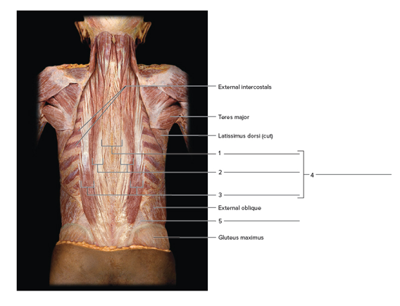
Label the deep back muscles of a cadaver, using the terms provided. The trapezius, latissimusdorsi, rhombus, and serratus posterior have been removed.

Terms:
Erector spinae
lliocostalis
Longissimus
Quadratuslumborum
Spinalis
Expert Solution & Answer
Want to see the full answer?
Check out a sample textbook solution
Students have asked these similar questions
Muscles of the Lower LimbUse the key terms to respond to the descriptions below. (Some terms may be used more than once.)Key:adductor groupbiceps femorisextensor digitorum longusfibularis (peroneus) musclesgastrocnemiusgluteus maximusrectus femorissemimembranosussemitendinosustibialis anteriortibialis posteriorvastus muscles
----------------------, 1. adduct the thigh, as when standing at attention----------------------, 2. extends the toes----------------------, 3. extends knee and flexes thigh--------------------, 4. used to extend the hip when climbing stairs
----------------------, 5. prime movers of plantar flexion (two muscles) of the foot
MUSCULOSKELETAL ANA
THIRD EDITIO
Antagonistic muscle action chart. Cervical and lumbar spine
Complete the chart by listing the muscle(s) or parts of muscles that are antagonist in their actions to the muscles
in the left column.
Antagonist
Agonist
Splenius capitis
Splenius cervicis
Sternocleidomastoid
Erector spinae
Rectus abdominis
External oblique abdominal
Internal oblique abdominal
Quadratus lumborum
itic
Match the muscles that are antagonistic in their actions to the muscles listed.
Adductor brevis
Adductor longus
Adductor magnus
Biceps femoris
Gluteus maximus
Gluteus medius…
Chapter 24 Solutions
Laboratory Manual For Human Anatomy & Physiology
Ch. 24 - The abdominal wall muscles possess the following...Ch. 24 - The abdominal muscle nearest the midline is the a....Ch. 24 - Which of the following muscles would be most...Ch. 24 - The four muscles of the abdominal wall a. contain...Ch. 24 - The iliocostalis represents the lateral muscle...Ch. 24 - The pelvic floor muscles are all arranged in a...Ch. 24 - Label the deep back muscles of a cadaver, using...Ch. 24 - Label the muscles of the abdominal wall.Ch. 24 - Name the abdominal muscles a surgeon would incise...Ch. 24 - Complete the following statements: 1. A band of...
Ch. 24 - Complete the following statements: 2. The...Ch. 24 - Complete the following statements: 3. The...Ch. 24 - Complete the following statements: 4. The action...Ch. 24 - Complete the following statements: 5. The action...Ch. 24 - Complete the following statements: 6. The...Ch. 24 - Complete the following statements: 7. The...Ch. 24 - Complete the following statements: 8. The origin...Ch. 24 - Complete the following statements: 9. The...Ch. 24 - Complete the following statements: 10. Name four...Ch. 24 - Complete the following statements: 11. The actions...Ch. 24 - Complete the following statements: 12. Name the...Ch. 24 - Complete the following statements: 1. The levator...Ch. 24 - Complete the following statements: 2. The...Ch. 24 - Complete the following statements: 3. The...Ch. 24 - Complete the following statements: 4. In females,...Ch. 24 - Prob. 3.5ACh. 24 - Complete the following statements: 6. In the...Ch. 24 - Complete the following statements: 7. The...Ch. 24 - Complete the following statements: 8. The...Ch. 24 - Complete the following statements: 9. The...Ch. 24 - Complete the following statements: 10. Name five...
Knowledge Booster
Learn more about
Need a deep-dive on the concept behind this application? Look no further. Learn more about this topic, biology and related others by exploring similar questions and additional content below.Similar questions
- Describe 1. The location including the origin, insertion and Function of each of the following muscles. Occipitofrontalis Buccinator Orbicularis oculi Orbicularis oris Masseter Temporalis Sternocleidomastoidarrow_forwardPut the right muscle name in the right boxarrow_forwardMatch the term with the correct description. these are the terms: Anterior longitudinal ligament (ALL) Posterior longitudinal ligament (PLL) Ligamentum flava Ligamentum nuchae these are the descriptions: This is a large ligament located between the posterior muscles of C1 to C6-C7 spinous processes. This ligament becomes part of the supraspinous ligament at C7. It limits hyperflexion of the neck. Supraspinous ligament This is a thick ligament connecting the spinous processes of C7 down to the L3 or L4 vertebrae. It joins other ligaments to limit flexion of the spinal column. This ligament is attached to the posterior surface of the vertebrae body and the intervertebral discs in the spinal canal. It starts from C2 and extends downward to the sacrum. It prevents hyperflexion. It also supports the spinal column. Located within the spinal canal, it is found on the posterior bodies of the vertebrae. It starts at C2 and moves downward to the sacrum. It prevents hyperflexion. It also supports…arrow_forward
- Label the dorsal muscles. Use the choices provided at the top-middle part of the image. Note that the asterisks indicate the frequency that the choice may be used. (e.g. ** - can be used twice). Specimen: frogarrow_forwardmatcharrow_forwardFor each of the muscles below, state the criteria used for naming the muscle. For example, serratus anterior is a saw-toothed anterior muscle. 1. Latissimus dorsi 2. Rectus abdominis 3. Trapezius 4. Biceps brachii 5. Levator scapulae 6. Flexor carpi radialis 7. Piriformis 8. Gluteus medius 9. Rhomboid major 10. Pectoralis majorarrow_forward
- Nonearrow_forwardEpicranial aponeurosis Frontal belly -Epicranius Corrugator supercilii- Occipital belly Orbicularis oculi- Levator labi superioris Temporalis Zygomaticus minor and major Buccinator Risorius Masseter Sternocleidomastoid Orbicularis oris - Mentalis- Trapezius Depressor labii inferioris Splenius capitis Depressor anguli oris- Platysma (a) The flexion of which one of the following muscles would cause you frown? O platysma O buccinator orbicularis oculi O occipitofrontalis orbicularis orisarrow_forwardDrag the correct label to the appropriate location to identify the muscles of the anterior thigh. III Sartorius Tliopsoas Rectus femoris Pectineus Adductor Vastus lateralis magnus Gracilis Tensor fascian Adductor longus latao Vastus medias Reset Helparrow_forward
arrow_back_ios
SEE MORE QUESTIONS
arrow_forward_ios
Recommended textbooks for you
 Fundamentals of Sectional Anatomy: An Imaging App...BiologyISBN:9781133960867Author:Denise L. LazoPublisher:Cengage Learning

Fundamentals of Sectional Anatomy: An Imaging App...
Biology
ISBN:9781133960867
Author:Denise L. Lazo
Publisher:Cengage Learning
Chapter 7 - Human Movement Science; Author: Dr. Jeff Williams;https://www.youtube.com/watch?v=LlqElkn4PA4;License: Standard youtube license